Case of primary low-grade neuroendocrine carcinoma of the skin
- PMID: 38110346
- PMCID: PMC10749125
- DOI: 10.1136/bcr-2023-257569
Case of primary low-grade neuroendocrine carcinoma of the skin
Abstract
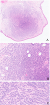
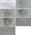
A man presents a 4 mm skin tumour at his general practitioner. The tumour is removed on the suspicion of a dermatofibroma. Important differential diagnoses are sebaceous neoplasms, melanomas, Merkel cell carcinomas and large cell neuroendocrine carcinoma, and metastases of neuroendocrine neoplasms from the gut or lung. Immunohistochemical staining excluded sebaceous neoplasm, melanoma and Merkel cell carcinoma, however, was positive for multiple neuroendocrine markers. Relevant scans showed no signs of a primary tumour anywhere else. The final diagnosis was a primary low-grade neuroendocrine carcinoma of the skin. At 30 months follow-up, there was no sign of recurrence.
Keywords: Dermatology; Neuroendocrinology; Pathology.
© BMJ Publishing Group Limited 2023. Re-use permitted under CC BY-NC. No commercial re-use. See rights and permissions. Published by BMJ.
Conflict of interest statement
Competing interests: GD received teaching activities for Ipsen, advanced Pharma and Sam Nordic.
Figures


Similar articles
-
Merkel cell carcinoma expresses K homology domain-containing protein overexpressed in cancer similar to other high-grade neuroendocrine carcinomas.Hum Pathol. 2009 Feb;40(2):238-43. doi: 10.1016/j.humpath.2008.07.009. Epub 2008 Oct 5. Hum Pathol. 2009. PMID: 18835627
-
Diagnostic accuracy of a panel of immunohistochemical and molecular markers to distinguish Merkel cell carcinoma from other neuroendocrine carcinomas.Mod Pathol. 2019 Apr;32(4):499-510. doi: 10.1038/s41379-018-0155-y. Epub 2018 Oct 22. Mod Pathol. 2019. PMID: 30349028
-
Differentiating Merkel cell carcinoma of lymph nodes without a detectable primary skin tumor from other metastatic neuroendocrine carcinomas: The ELECTHIP criteria.J Am Acad Dermatol. 2018 May;78(5):964-972.e3. doi: 10.1016/j.jaad.2017.11.037. Epub 2017 Nov 24. J Am Acad Dermatol. 2018. PMID: 29180096
-
[Merkel cell carcinoma].Hautarzt. 2002 Oct;53(10):652-8. doi: 10.1007/s00105-001-0318-4. Hautarzt. 2002. PMID: 12297946 Review. German.
-
A Merkel cell carcinoma presenting as a solitary lymph node metastasis without a primary lesion. Report of a case and review of the literature.Acta Chir Belg. 2012 Jul-Aug;112(4):317-21. Acta Chir Belg. 2012. PMID: 23009000 Review.
Cited by
-
Sweat gland carcinoma with neuroendocrine differentiation of the scalp with elevated serum chromogranin A levels.JAAD Case Rep. 2025 Jan 28;60:41-43. doi: 10.1016/j.jdcr.2024.12.002. eCollection 2025 Jun. JAAD Case Rep. 2025. PMID: 40353101 Free PMC article. No abstract available.
References
Publication types
MeSH terms
Substances
LinkOut - more resources
Full Text Sources
Medical
