. 2023 Dec 18;14(1):8006.
doi: 10.1038/s41467-023-43730-0.

Genetic and epigenetic features of bilateral Wilms tumor predisposition in patients from the Children's Oncology Group AREN18B5-Q

Affiliations

Genetic and epigenetic features of bilateral Wilms tumor predisposition in patients from the Children's Oncology Group AREN18B5-Q

Andrew J Murphy et al. Nat Commun. .

Abstract

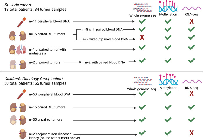
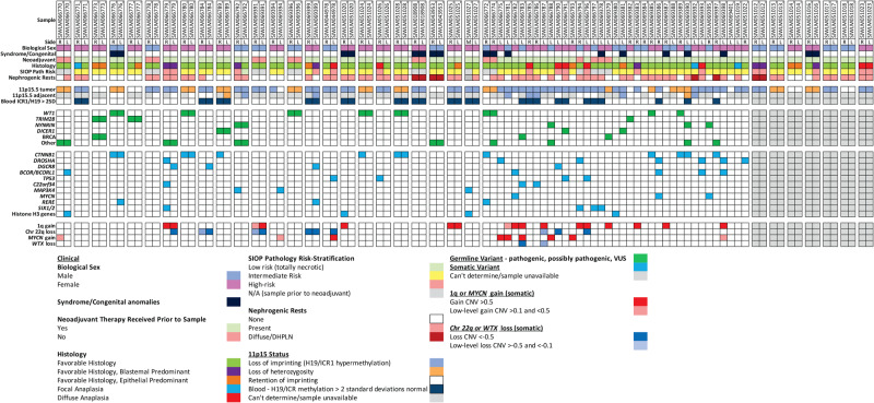
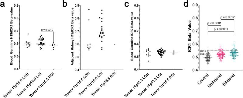
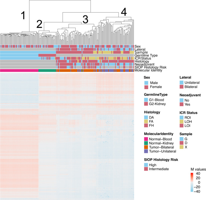
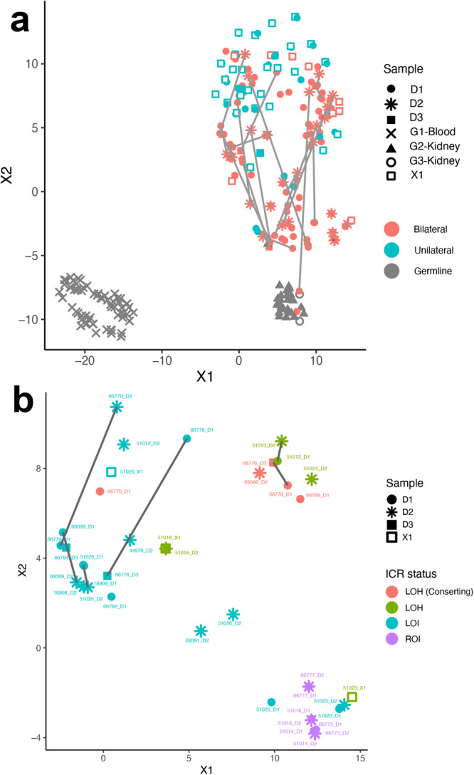
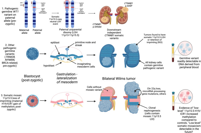
Developing synchronous bilateral Wilms tumor suggests an underlying (epi)genetic predisposition. Here, we evaluate this predisposition in 68 patients using whole exome or genome sequencing (n = 85 tumors from 61 patients with matched germline blood DNA), RNA-seq (n = 99 tumors), and DNA methylation analysis (n = 61 peripheral blood, n = 29 non-diseased kidney, n = 99 tumors). We determine the predominant events for bilateral Wilms tumor predisposition: 1)pre-zygotic germline genetic variants readily detectable in blood DNA [WT1 (14.8%), NYNRIN (6.6%), TRIM28 (5%), and BRCA-related genes (5%)] or 2)post-zygotic epigenetic hypermethylation at 11p15.5 H19/ICR1 that may require analysis of multiple tissue types for diagnosis. Of 99 total tumor specimens, 16 (16.1%) have 11p15.5 normal retention of imprinting, 25 (25.2%) have 11p15.5 copy neutral loss of heterozygosity, and 58 (58.6%) have 11p15.5 H19/ICR1 epigenetic hypermethylation (loss of imprinting). Here, we ascertain the epigenetic and genetic modes of bilateral Wilms tumor predisposition.


Conflict of interest statement

The authors declare no competing interests.

Figures

Fig. 1
Fig. 1. Specimens and molecular assays used in the current study.
Whole exome or whole genome sequencing germline variant calls were made using DNA obtained from 61 patients with available DNA from peripheral blood (n = 11 SJCRH and n = 50 COG). Paired tumor sets are samples from both right and left tumors in a patient with synchronous BWT (n = 30). Unpaired tumors are samples from either the right or left tumor in a patient with synchronous BWT, but for whom one side was not available for analysis (n = 37). Adjacent non-diseased kidney was confirmed by a pathologist and came from patients with tumors in the study. Abbreviations: R – right L – left; Seq -sequencing. Graphic made with biorender.com.
Fig. 2
Fig. 2. Spectrum of clinical, genetic, and epigenetic features associated with bilateral Wilms tumor.
Paired synchronous BWT (n = 23; n = 1 primary tumor + corresponding metastasis) are outlined in paired boxes on the left side of the graphic. Additional paired synchronous BWT (n = 7) that did not have a germline peripheral blood sample available are shown in paired boxes on the right side of the graphic. These 7 paired synchronous BWT specimens were analyzed for methylation and RNA-seq only. Gray boxes indicate when a given finding could not be assessed due to sample availability. Unpaired specimens from BWT patients are outlined in the adjacent boxes without spacing in the center of the graphic. R right, L left, Adjacent adjacent non-diseased kidney, SD standard deviation, Chr chromosome, DHPLN diffuse hyperplastic perilobar nephroblastomatosis, CNV chromosomal copy number variant. Source data are available in Supplementary Data Files 1, 2 and 5.
Fig. 3
Fig. 3. Exploratory analysis of low-level H19/ICR1 hypermethylation detectable in peripheral blood.
a Patients from the current study COG cohort with BWT containing 11p15.5 LOI were found to have a statistically significant increase in 11p15.5 H19/ICR1 methylation detected in the peripheral blood when compared to patients with tumors having 11p15.5 retention of imprinting (unpaired, two-tailed t test p = 0.0210; n = 50 biologically independent samples). b 11p15.5 LOI was often detectable above the threshold value (β > 0.7) for loss of imprinting in adjacent non-diseased kidney tissues (n = 29 biologically independent samples). c No differences were detected among patient groups at 11p15.5 KCNQ1OT1/ICR2 in peripheral blood (n = 50 biologically independent samples). Of note, the single sample outlier with tumor 11p15.5 LOH, hypermethylation at H19/ICR1, and hypomethylation at KCNQ1OT1/ICR2 detected in the peripheral blood was confirmed to have mosaicism for 11p15.5 LOH detected in peripheral blood by whole genome sequencing. For (ac), lines represent median values. d A significant increase in H19/ICR1 methylation detected in peripheral blood was noted when BWT patients (n = 78) were compared to unilateral WT (n = 154 biologically independent samples) and healthy community control subjects (n = 282 biologically independent samples; p values are ordinary one-way ANOVA with pairwise values corrected for multiple comparisons). 26 BWT patient blood samples (green triangles; n = 20 biologically independent from current study, n = 6 from survivor cohort) had low-level H19/ICR1 gain of methylation defined as a β value greater than two standard deviations (2 SD) above the mean from the healthy community control cohort. For boxes in (d), measure of center lines are mean and whiskers are SD. LOH loss of heterozygosity, LOI loss of imprinting. Source data are available in the Source Data File.
Fig. 4
Fig. 4. Unsupervised hierarchical clustering of methylation M-values from the top 10,000 most variable probes in the 850 K EPIC Methylation Beadchip array.
Distinct clustering of DNA samples derived from peripheral blood (cluster 1), adjacent non-diseased kidney (cluster 2), a cluster of predominantly BWT (cluster 3), and a cluster of predominantly unilateral WT (cluster 4). Within the BWT cluster (cluster 3), samples with 11p15.5 LOH, 11p15.5 LOI, and 11p15.5 ROI cluster together. Notably, the BWT cluster (cluster 3) joins with adjacent non-diseased kidney (cluster 2) more closely than the unilateral WT cluster. N = 212 biologically independent samples. Histology – DA – diffuse anaplasia, FA focal anaplasia, FH favorable histology. ICR status: ROI retention of imprinting, LOH loss of heterozygosity, LOI loss of imprinting. Tumor type: G – germline sample, D – primary tumor sample, X – patient-derived xenograft. Source data are available in the Source Data File.
Fig. 5
Fig. 5. TSNE clustering analysis of 850 K EPIC Methylation Beadchip array.
a Predominant clusters of unilateral (teal) and bilateral (salmon) WT. BWT cluster closer to adjacent non-diseased kidney (gray triangles) than unilateral WT. Gray lines connect synchronous BWT. However, large differences in synchronous BWT correlated with differential tumor purity in each DNA sample. N = 267 biologically independent samples. b Clustering of BWT with a tumor purity filter applied (excluding specimens with tumor purity <80%) shows near adjacent clustering of paired synchronous BWT specimens. Within BWT, samples cluster according to 11p15.5 status: 11p15.5 LOI (teal), 11p15.5 LOH (red and green), and 11p15.5 ROI (purple). The greatest difference between paired synchronous BWT samples is seen in samples with 11p15.5 LOI (samples connected by gray lines). N = 40 biologically independent samples. G1 blood-derived germline DNA, G2 and G3 adjacent non-diseased kidney derived DNA, D1-D3 tumor derived DNA, X1 patient-derived xenograft DNA, ICR status – LOH – loss of heterozygosity, LOI loss of imprinting, ROI retention of imprinting. Source Data are available in the Source Data File.
Fig. 6
Fig. 6. Summary of Proposed Mechanisms for Bilateral Wilms tumor development.
The top panel depicts the molecular sequence leading to BWT in patients with pathogenic heterozygous WT1 germline variants. Here, somatic 11p15.5 copy neutral loss of heterozygosity causes biallelic inactivation of WT1 and biallelic expression of IGF2, a sequence which is often followed by downstream distinct CTNNB1 somatic variants unique to each tumor. The middle panel depicts the general sequence of BWT development due to a pre-zygotic pathogenic germline variant in which the germline variant is present in all kidney cells. The bottom panel depicts somatic mosaic 11p15.5 loss of imprinting, in which 11p15.5 H19/ICR1 gain of methylation occurs on the maternal allele in a post-zygotic embryonic cell. This event must occur prior to lateralization of cells fated to become mesoderm during embryonic gastrulation. At this time in embryonic development, the cells that give rise to the intermediate mesoderm and therefore the kidneys are anatomically sequestered from one another (right and left). This lateralization results in a somatic mosaic distribution of 11p15.5 LOI throughout the body/mesoderm. Expansion of cellular clones containing the somatic mosaic alteration is termed clonal nephrogenesis and explains the detection of 11p15.5 LOI in adjacent non-diseased kidney tissue. BWT and/or multifocal WT arise from these clonal populations of kidney cells following additional somatic events including LOH of chromosome 22q, microRNA processing gene pathogenic variants, and others. LOH loss of heterozygosity, LOI loss of imprinting. Graphic made with biorender.com.

Update of

Similar articles

Cited by

References

    1. Davidoff AM. Wilms tumor. Adv. Pediatr. 2012;59:247–67. doi: 10.1016/j.yapd.2012.04.001. - DOI - PMC - PubMed
    1. Knudson AG, Strong LC. Mutation and cancer: A model for Wilms’ tumor of the kidney. J. Natl. Cancer Inst. 1972;48:313–24. - PubMed
    1. Spreafico F, et al. Wilms tumour. Nat. Rev. Dis. Prim. 2021;7:75. doi: 10.1038/s41572-021-00308-8. - DOI - PubMed
    1. Beckwith JB, Kiviat NB, Bonadio JF. Nephrogenic rests, nephroblastomatosis, and the pathogenesis of Wilms’ tumor. Pediatr. Pathol. 1990;10:1–36. doi: 10.3109/15513819009067094. - DOI - PubMed
    1. Chernin G, et al. Genotype/phenotype correlation in nephrotic syndrome caused by WT1 mutations. Clin. J. Am. Soc. Nephrol. 2010;5:1655–62. doi: 10.2215/CJN.09351209. - DOI - PMC - PubMed

Publication types

Associated data