. 2023 Dec 19;14(1):8423.
doi: 10.1038/s41467-023-44152-8.

Recently activated CD4 T cells in tuberculosis express OX40 as a target for host-directed immunotherapy

Affiliations

Recently activated CD4 T cells in tuberculosis express OX40 as a target for host-directed immunotherapy

Abigail R Gress et al. Nat Commun. .

Abstract

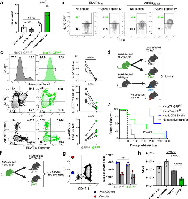
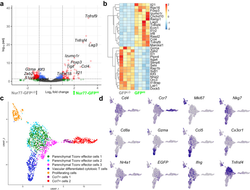
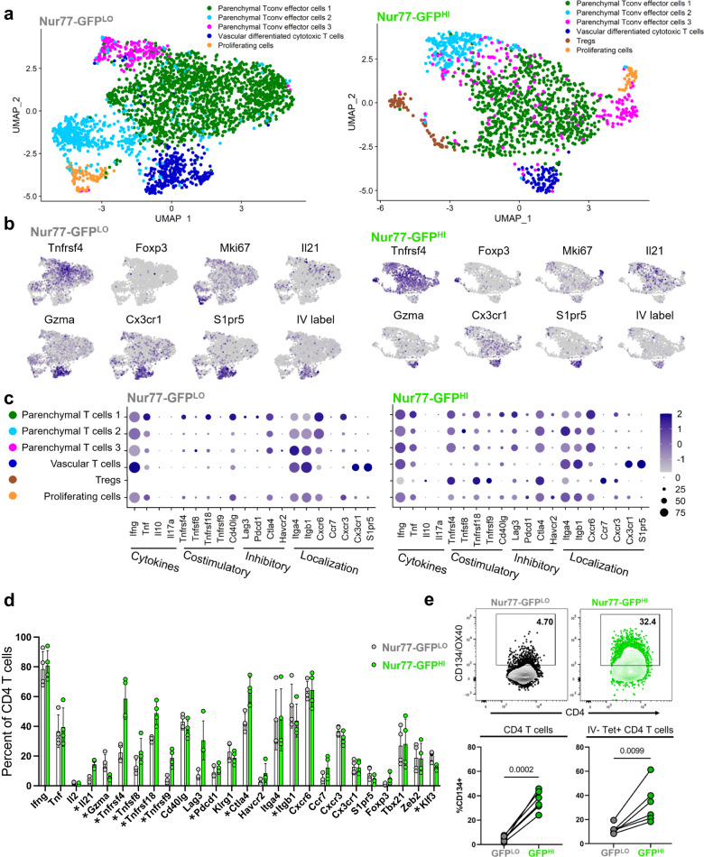
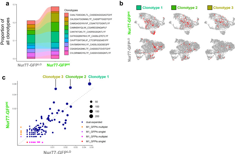
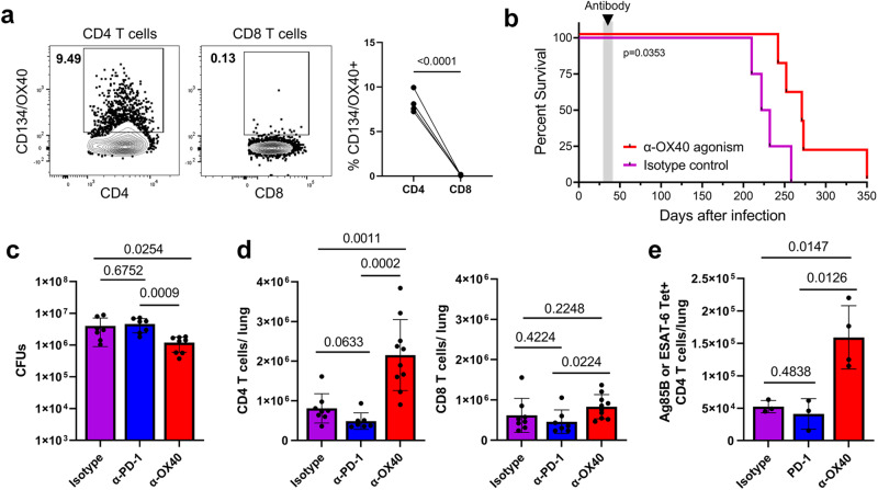
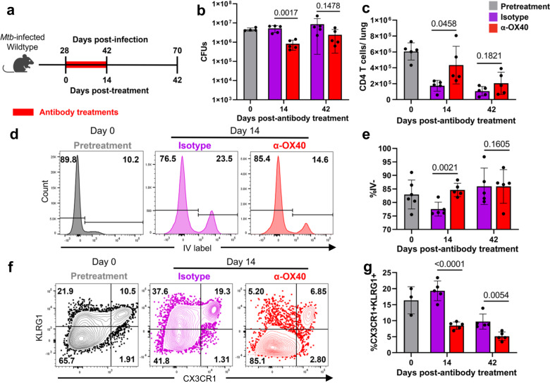
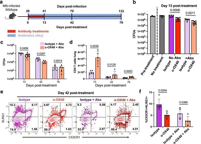
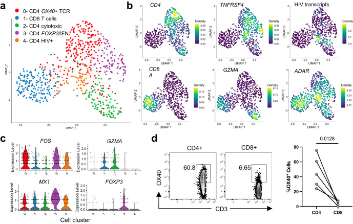
After Mycobacterium tuberculosis (Mtb) infection, many effector T cells traffic to the lungs, but few become activated. Here we use an antigen receptor reporter mouse (Nur77-GFP) to identify recently activated CD4 T cells in the lungs. These Nur77-GFPHI cells contain expanded TCR clonotypes, have elevated expression of co-stimulatory genes such as Tnfrsf4/OX40, and are functionally more protective than Nur77-GFPLO cells. By contrast, Nur77-GFPLO cells express markers of terminal exhaustion and cytotoxicity, and the trafficking receptor S1pr5, associated with vascular localization. A short course of immunotherapy targeting OX40+ cells transiently expands CD4 T cell numbers and shifts their phenotype towards parenchymal protective cells. Moreover, OX40 agonist immunotherapy decreases the lung bacterial burden and extends host survival, offering an additive benefit to antibiotics. CD4 T cells from the cerebrospinal fluid of humans with HIV-associated tuberculous meningitis commonly express surface OX40 protein, while CD8 T cells do not. Our data thus propose OX40 as a marker of recently activated CD4 T cells at the infection site and a potential target for immunotherapy in tuberculosis.


Conflict of interest statement

The authors declare no competing interests.

Figures

Fig. 1
Fig. 1. Nur77-GFPHI and Nur77-GFPLO CD4 T cells are phenotypically and functionally distinct.
a Frequency of Nur77-GFPHI CD4 T cells in lungs of Nur77-GFP or wildtype mice at four weeks post-infection or uninfected. Gated on live CD4+ CD44+ Nur77-GFPHI cells. P values calculated with an unpaired two-tailed t-test, 3 mice per group. b Flow cytometry of Ag85B- or ESAT-6-specific CD4 T cells from Nur77-GFP lungs harvested at four weeks post-infection. Mice indicated received Ag85B peptide injected six hours before harvest. Gated on live CD3+ CD4+ CD44+ Tetramer+ cells. Tetramer+ cells represent either Ag85B or ESAT-6 specificity. Representative plots from experiment with 4 mice per group. c Flow cytometry of Nur77-GFPLO and Nur77-GFPHI CD4 T cells from Nur77-GFP lungs harvested at four weeks post-infection. CD45.2 fluorescent antibody injected intravenously immediately before harvest identifies vascular cells. Gated on live CD3+ CD4+ CD44+ and either lowest or highest 1/3 of Nur77-GFP expression. Further gated on IV-, CX3CR1+ KLRG1+, or Tetramer+ cells. Tetramer+ cells represent either Ag85B or ESAT-6 specificity. P values calculated with a paired two tailed t-test, 4 or 5 mice per group. d Nur77-GFPLO and Nur77-GFPHI CD4 T cells sorted from Nur77-GFP lungs at four weeks post-infection and injected into one week post-infection TCRα-/- mice. Donor cells sorted for live CD3+ CD4+ CD44+ Nur77-GFPLO, Nur77-GFPHI, or total bulk CD4 T cells. e Curves show survival of recipient TCRα-/- mice. P value calculated with a two-sided Mantel-Cox test, 4-6 mice per group. f–h Nur77-GFPLO and Nur77-GFPHI CD4 T cells sorted from Nur77-GFP lungs at four weeks post-infection and injected into infection-matched wildtype CD45.1 mice. Donor cells sorted on live CD4+ CD44+ Nur77-GFPLO or Nur77-GFPHI cells. Recipient and no transfer wildtype CD45.1 lungs harvested at one week post-transfer for flow cytometry and lung bacterial load. g Number and lung localization of donor and recipient cells at one-week post-transfer. CD45.2 fluorescent antibody injected immediately before harvest identifies vascular cells. Gated on live CD4+ CD44+ cells. Cell numbers assessed using counting beads. P value calculated with an unpaired two tailed t-test. Mice per group: 4 for Nur77-GFPLO recipients or 3 for Nur77-GFPHI. h Lung bacterial load at one-week post-transfer or without transfer. Pre-transfer wildtype CD45.1 lungs harvested at four weeks post-infection. P values calculated with an unpaired two-tailed t-test. Mice per group: 5 for pre-transfer, 4 for no transfer, 4 for Nur77-GFPLO recipients or 3 for Nur77-GFPHI. Error bars indicate standard deviation.
Fig. 2
Fig. 2. Mtb-specific CD4 T cells include diverse phenotypes.
a, b Differential gene expression analysis of Nur77-GFPLO and Nur77-GFPHI CD4 T cells sorted from lungs at four weeks post-infection. Data representative of 4 biological replicates. Volcano plot highlights genes in red with log2 fold change > 1 and p value based on the two-tailed Wald test adjusted for multiple hypothesis testing using Benjamini-Hochberg correction with cutoff of -log padj > 1. Heatmap shows scaled differential expression between Nur77-GFPLO and Nur77-GFPHI cells of a selection of genes with -log padj > 1, with same statistical approach as above. Source data for a,b are located in Supplementary Data S1. c UMAP projection of single-cell transcriptomes of 1,795 cells from the lungs of Nur77-GFP mice at four weeks post-infection, that underwent magnetic CD4 isolation and tetramer enrichment for Ag85B- and ESAT-6-specificity. Data represents cells pooled from 3 individual mice. Colors indicate transcriptomic clusters with manual annotation based on differential expression of marker genes. d Feature plots show regions of elevated expression of indicated genes. Source data for c, d located in Supplementary Data S2.
Fig. 3
Fig. 3. Nur77-GFPHI and Nur77-GFPLO CD4 T cells differ in expression of effector molecules.
a–d Single-cell RNA-Sequencing of Nur77-GFPLO and Nur77-GFPHI CD4 T cells (2,664 and 1386, respectively) sorted from Nur77-GFP lungs at four weeks post-infection. a Data show paired cells from an individual mouse, representative of 4 biological replicates. Colors indicate transcriptomic clusters with manual annotation based on differential expression of marker genes. b Feature plots show regions of elevated expression of indicated genes in Nur77-GFPLO and Nur77-GFPHI cell populations. For IV label, CD45.2 CITE-seq antibody was injected immediately before harvest. c Dot plots show relative abundance (color scale) and prevalence (circle size) of indicated gene expression in each cluster, in Nur77-GFPLO and Nur77-GFPHI cell populations. Source data for a–c are located in Supplementary Data S3d Columns indicate percent of Nur77-GFPLO and Nur77-GFPHI CD4 T cells expressing each gene, paired from 4 individual mice. Asterisks indicate q value < 0.05 for paired two-tailed t tests adjusted for multiple comparisons, with precise q value listed in Supplementary Data S4. e Flow cytometry of OX40 surface protein expression on Nur77-GFPLO and Nur77-GFPHI CD4 T cells from Nur77-GFP lungs harvested at four weeks post-infection. CD45.2 fluorescent antibody injected immediately before harvest identifies vascular cells. Tetramer+ cells represent either Ag85B or ESAT-6 specificity. Gated on IV- Tetramer+ or bulk live CD4+ CD44+ Nur77-GFPLO or Nur77-GFPHI cells. P values calculated with a paired two-tailed t-test, 5-6 mice per group. Error bars indicate standard deviation.
Fig. 4
Fig. 4. Nur77-GFPHI and Nur77-GFPLO CD4 T cells differ in expression of TCR clonotypes.
a Relative abundance of top 10 clonotypes from single-cell TCR-Sequencing of Nur77-GFPLO and Nur77-GFPHI CD4 T cells sorted from the lungs of one Nur77-GFP mouse at four weeks post-infection. Samples sorted on live CD3+CD4+CD44+ Nur77-GFPLO or Nur77-GFPHI cells. Amino acid sequences represent TCR CDR3 regions expressed as “TCRα_TCRβ“. b Feature plots show the localization of three expanded TCR clonotypes on gene expression UMAPs from Nur77-GFPLO and Nur77-GFPHI CD4 T cells. c Scatter plot shows the prevalence of TCR clonotypes among Nur77-GFPLO vs. Nur77-GFPHI cells. Blue dots indicate expanded clonotypes. Circle size indicates total abundance of each expanded clonotype.
Fig. 5
Fig. 5. OX40 agonism improves TB outcomes and increases CD4 T cell numbers.
a Flow cytometry of CD4 and CD8 T cells from wildtype C57BL/6 lungs at four weeks post-infection. Gated on live CD4+cCD44+ or CD8+cCD44+ cells. P value = 0.00007 calculated with a paired two-tailed t-test, 4 mice per group. b–e Wildtype C57BL/6 mice at four weeks post-infection treated with 100 μg α-OX40 agonist, α-PD-1 blockade, or isotype control antibody injections twice weekly for two weeks. Mice followed for survival or lungs harvested one day after the last antibody treatment for flow cytometry and lung bacterial load. b Curves show survival of α-OX40 or isotype control antibody-treated wildtype or TCRα-/- mice. P value calculated with a two sided Mantel-Cox test, 4-5 mice per group. c Lung bacterial load of α-OX40, α-PD-1, or isotype control antibody-treated mice. One-way ANOVA p = 0.0078; p values shown in figure calculated with post hoc testing adjusted for multiple comparisons. Mice per group: 6 for isotype, 7 for PD-1, and 8 for OX40. d Number of CD4 and CD8 T cells in the lungs of α-OX40, α-PD-1, or isotype control antibody treated mice. Gated on live CD4+ CD44+ or CD8+ CD44+ cells. Cell numbers assessed using counting beads. Data representative of two repeat experiments. One-way ANOVA for CD4 T cells p = 0.00002, CD8 T cells p = 0.1023; p values shown in figure calculated with post hoc testing adjusted for multiple comparisons. P value for PD-1 vs OX40 group = 0.00005. Mice per group: 8 for isotype, 7 for PD-1, and 10 for OX40. e Number of Tetramer+ CD4 T cells in the lungs of α-OX40, α-PD-1, or isotype control antibody-treated mice. Gated on live CD4+ CD44+ Tetramer+ cells. Tetramer+ cells represent either Ag85B or ESAT-6 specificity. Cell numbers assessed using counting beads. One-way ANOVA p = 0.0047, p values shown in figure adjusted for multiple comparisons. Mice per group: 3 for isotype, 3 for PD-1, and Mice per group: 8 for isotype, 7 for PD-1, and 10 for OX40. 4 for OX40. Error bars indicate standard deviation.
Fig. 6
Fig. 6. OX40 agonism alters CD4 T cell phenotype and retention at late timepoints.
a–g Wildtype C57BL/6 mice at four weeks post-infection treated with 100 μg α-OX40 agonist or isotype control antibody injections twice weekly for two weeks. Mice harvested at 0, 14, and 14 days post-antibody treatment for flow cytometry or lung bacterial load. b Lung bacterial load of α-OX40 or isotype control antibody treated mice. P values calculated with an unpaired two tailed t-test, adjusted for multiple comparisons. Mice per group: 4 for day 0, 5 each for Isotype or OX40 at day 14, and 5 each for Isotype or OX40 at day 42. c Number of CD4 T cells in the lungs of α-OX40 or isotype control antibody treated mice. Gated on live CD4+ CD44+ cells. Cell numbers assessed using counting beads. P values calculated with an unpaired two-tailed t-test, adjusted for multiple comparisons, 5 mice per group. d, e Frequency of parenchymal CD4 T cells in the lungs of α-OX40 or isotype control antibody-treated mice. CD45.2 fluorescent antibody injected immediately before harvest identifies vascular cells. Gated on live CD4+ CD44+ IV- cells. P values calculated with an unpaired two-tailed t-test, adjusted for multiple comparisons. Mice per group: 6 for day 0, 5 each for Isotype or OX40 at day 14, and 5 each for Isotype or OX40 at day 42. f, g Frequency of terminally differentiated CD4 T cells in the lungs of α-OX40 or isotype control antibody-treated mice. Gated on live CD4+ CD44+ CX3CR1+ KLRG1+ cells. P values calculated with an unpaired two-tailed t-test, adjusted for multiple comparisons. Mice per group: 3 for day 0, 5 each for Isotype or OX40 at day 14, and 5 each for Isotype or OX40 at day 42. Error bars indicate standard deviation.
Fig. 7
Fig. 7. OX40 agonism improves antibiotic treatment outcomes.
a–f Wildtype C57BL/6 mice at four weeks post-infection treated with 100 μg α-OX40 agonist or isotype control antibody injections twice weekly for two weeks. Antibiotic mice also treated from four weeks post-infection to harvest with isoniazid and rifampin in water continuously. Mice harvested at 0, 13, 42, and 76 days post-treatment for flow cytometry of lung bacterial load. b Lung bacterial load of α-OX40 antibody or isotype control antibody, with or without antibiotics. Pretreatment wildtype lungs harvested at 0 days post-treatment. Data representative of two repeat experiments. Two-way ANOVA results: antibiotic treatment contributed 43% of variance p < 0.0001, antibody treatment contributed 30% of variance p < 0.0001, interaction contributed 13% of variance p = 0.0015; p values shown in figure calculated with two-tailed t testing adjusted for multiple comparisons. Mice per group: 4 for pre-treatment, 5 for no treatment, 5 for no antibiotics + Isotype, 9 for no antibiotics + OX40, 5 for antibiotics + Isotype, 9 for antibiotics + OX40. c, d CFUs and number of CD4 T cells in the lungs of α-OX40 antibody or isotype control antibody treated mice. Gated on live CD4+ CD44+ cells. Cell numbers assessed using counting beads. P values calculated with an unpaired two tailed t-test, adjusted for multiple comparisons. Mice per group: 5 for isotype and 10 for OX40 at day 13, 5 each for isotype and OX40 at day 42, and 6 each for isotype and OX40 at day. e, f Frequency of terminally differentiated CD4 T cells in the lungs of α-OX40 antibody or isotype control antibody, with or without antibiotics. Gated on live CD4+ CD44+ CX3CR1+ KLRG1+ cells. Two-way ANOVA results: antibiotic treatment contributed 39% of variance p = 0.0005, antibody treatment contributed 27% of variance p = 0.0005, interaction contributed 1% of variance p = 0.43; p values shown in figure calculated with two-tailed t testing adjusted for multiple comparisons, 5 mice per group. Error bars indicate standard deviation.
Fig. 8
Fig. 8. Human CD4 T cells at the site of HIV-associated TB meningitis express OX40.
a UMAP representing single-cell RNA-Sequencing of CD4+ and CD8+ T cells sorted from cerebrospinal fluid (CSF) cells from a human person with HIV-associated TB meningitis. Sorted cells were gated on live, CD3+, CD4+ and CD8+ cells. Clusters were manually identified according to the differential expression of key genes, indicated as follows. b Plots show the relative density of cells expressing each marker gene. c Violin plots show each gene’s relative expression level by cells assigned to each phenotypic cluster. Source data for a–c are located in Supplementary Data S5. d OX40 surface expression in cells from the CSF of 5 individuals treated for HIV-associated TB meningitis. Flow cytometry dot plots indicate the percentage of CD4+ or CD8+ T cells from a single individual that express OX40, while column dot plots show the paired percentage of CD4 or CD8 T cells from 4 individuals. P value calculated with a paired two-tailed t-test.

References

    1. World Health Organization. GLOBAL TUBERCULOSIS REPORT 2021. (2021).
    1. Dartois VA, Rubin EJ. Anti-tuberculosis treatment strategies and drug development: challenges and priorities. Nat. Rev. Microbiol. 2022;20:685–701. doi: 10.1038/s41579-022-00731-y. - DOI - PMC - PubMed
    1. Leveton C, et al. T-Cell-Mediated Protection of Mice against Virulent Mycobacterium tuberculosis. Infect. Immun. 1989;57:390–395. doi: 10.1128/iai.57.2.390-395.1989. - DOI - PMC - PubMed
    1. Mogues T, Goodrich ME, Ryan L, LaCourse R, North RJ. The relative importance of T cell subsets in immunity and immunopathology of airborne Mycobacterium tuberculosis infection in mice. J. Exp. Med. 2001;193:271–280. doi: 10.1084/jem.193.3.271. - DOI - PMC - PubMed
    1. Chamie G, et al. Significant variation in presentation of pulmonary tuberculosis across a high resolution of CD4 strata. Int. J. Tuberc. Lung Dis. 2010;14:1295–1302. - PMC - PubMed

Publication types

Substances