. 2023 Dec 18;13(1):22558.
doi: 10.1038/s41598-023-49874-9.

Fenofibrate reduces glucose-induced barrier dysfunction in feline enteroids

Affiliations

Fenofibrate reduces glucose-induced barrier dysfunction in feline enteroids

Charles K Crawford et al. Sci Rep. .

Abstract

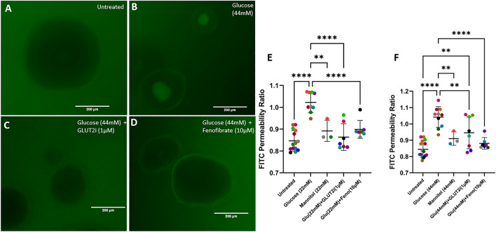
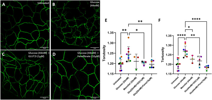
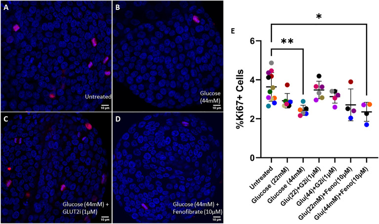
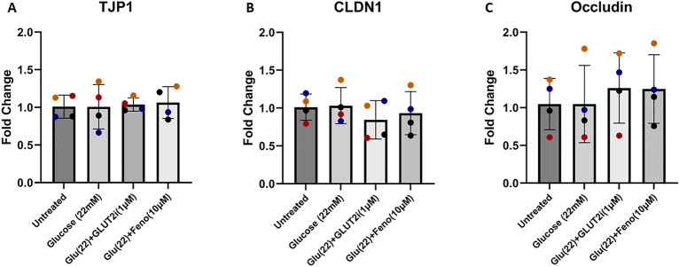
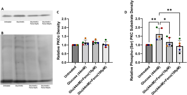
Diabetes mellitus (DM) is a common chronic metabolic disease in humans and household cats that is characterized by persistent hyperglycemia. DM is associated with dysfunction of the intestinal barrier. This barrier is comprised of an epithelial monolayer that contains a network of tight junctions that adjoin cells and regulate paracellular movement of water and solutes. The mechanisms driving DM-associated barrier dysfunction are multifaceted, and the direct effects of hyperglycemia on the epithelium are poorly understood. Preliminary data suggest that fenofibrate, An FDA-approved peroxisome proliferator-activated receptor-alpha (PPARα) agonist drug attenuates intestinal barrier dysfunction in dogs with experimentally-induced DM. We investigated the effects of hyperglycemia-like conditions and fenofibrate treatment on epithelial barrier function using feline intestinal organoids. We hypothesized that glucose treatment directly increases barrier permeability and alters tight junction morphology, and that fenofibrate administration can ameliorate these deleterious effects. We show that hyperglycemia-like conditions directly increase intestinal epithelial permeability, which is mitigated by fenofibrate. Moreover, increased permeability is caused by disruption of tight junctions, as evident by increased junctional tortuosity. Finally, we found that increased junctional tortuosity and barrier permeability in hyperglycemic conditions were associated with increased protein kinase C-α (PKCα) activity, and that fenofibrate treatment restored PKCα activity to baseline levels. We conclude that hyperglycemia directly induces barrier dysfunction by disrupting tight junction structure, a process that is mitigated by fenofibrate. We further propose that counteracting modulation of PKCα activation by increased intracellular glucose levels and fenofibrate is a key candidate regulatory pathway of tight junction structure and epithelial permeability.


Conflict of interest statement

The authors declare no competing interests.

Figures

Figure 1
Figure 1
Fenofibrate treatment prevents the increase in barrier permeability that is induced by supraphysiologic glucose concentration. (AD) Representative images of feline enteroids following exposure to 4 kDa FITC-Dextran following 24 h treatment with control media (A), 44 mM added glucose (B), 44 mM added glucose with GLUT2i (C), and 44 mM added glucose and fenofibrate (D). Images are acquired with a GFP LED light cube (470/525 nm ex/em). Scale bars denote 200 µm. (EF) Luminal FITC intensity normalized to basolateral FITC intensity following 24 h treatment with glucose, mannitol, glucose with GLUT2-selective inhibitor, or glucose with fenofibrate. One-way ANOVA (α = 0.05) with Holm-Šídák post hoc analysis determined that treatment with 22 mM or 44 mM additional glucose significantly increased barrier permeability (p < 0.0001 for both treatments). Mannitol treatment significantly reduced enteroid permeability compared to matching concentration of glucose treatment for both 22 mM and 44 mM treatements (p = 0.0012 and p = 0.0097, respectively). Enteroids treated with glucose and 1 µM GLUT2-selective inhibitor displayed significantly reduced barrier permeability compared to those treated with only glucose for both 22 mM and 44 mM treatments (p < 0.0001 and p = 0.0097, respectively), but co-treatment with GLUT2-selective inhibitor did increase barrier permeability relative to untreated enteroids for 44 mM glucose-treated enteroids (p = 0.0034). Enteroids treated with glucose and 10 µM fenofibrate displayed significantly reduced permeability compared to those treated with only glucose for both 22 mM and 44 mM glucose treatments (p < 0.0001 for both treatments). Data are presented as mean ± SD with each point representing an experimental mean. Colors represent individual experiments from one organoid line.
Figure 2
Figure 2
Fenofibrate treatment prevents the increase in tight junction tortuosity that is induced by supraphysiologic glucose concentration. (AD) Representative ZO-1 (Alexa Fluor 488) staining of feline enteroids following 24 h treatment with control media (A), 44 mM added glucose (B), 44 mM added glucose with GLUT2i (C), and 44 mM added glucose and fenofibrate (D). Scale bars denote 10 µm. (EF) Quantification of ZO-1 tortuosity in experimental enteroids. Treatment with 22 mM or 44 mM glucose significantly increased ZO-1 tortuosity (p = 0.0023, p < 0.0001, respectively) as determined by one-way ANOVA (α = 0.05) and Holm-Šídák post hoc analysis. 44 mM mannitol treatment significantly decreased junctional tortuosity compared to treatment with an equal concentration of glucose (p = 0.0416). Enteroids treated with 22 mM or 44 mM glucose and 1 µM GLUT2-selective inhibitor displayed significantly reduced ZO-1 tortuosity compared to those treated with only glucose (p = 0.0369 and p = 0.0020, respectively). Enteroids treated with 10 µM fenofibrate in addition to 22 mM or 44 mM glucose displayed significantly reduced ZO-1 tortuosity compared to those treated with only glucose (p = 0.0053, p < 0.0001, respectively). Data are presented as mean ± SD with each point representing an experimental mean. Colors represent individual experiments from one organoid line.
Figure 3
Figure 3
Supraphysiologic glucose concentration reduces cellular proliferation in feline enteroids. (AD) Representative confocal images of feline enteroids stained for Ki67 (Alexa Fluor 594) and DAPI following 24 h treatment with control media (A), 44 mM added glucose (B), 44 mM added glucose with GLUT2i (C), and 44 mM added glucose and fenofibrate (D). Scale bars denote 10 µm. (E) Percentage of proliferating cells measured by confocal microscopy. Treatment with 44 mM glucose with or without fenofibrate induced a significant reduction in proliferation compared to untreated enteroids (p = 0.0032 without fenofibrate, p = 0.0246 with fenofibrate) as determined by Kruskal–Wallis non-parametric ANOVA (α = 0.05) with Dunn’s post hoc analysis. Data are presented as mean ± SD with each point representing an experimental mean. Colors represent individual experiments from one organoid line.
Figure 4
Figure 4
Supraphysiologic glucose concentration does not alter apoptotic rate in feline enteroids. (AD) Representative confocal images of feline enteroids stained for Cleaved Caspase-3 (Alexa Fluor 594) and DAPI following 24 h treatment with control media (A), 44 mM added glucose (B), 44 mM added glucose with GLUT2i (C), and added 44 mM glucose and fenofibrate (D). Scale bars denote 10 µm. (E) Fluorescence intensity of Cleaved Caspase-3 normalized to DAPI measured by confocal microscopy. No significant effect of treatment was detected by Kruskal–Wallis non-parametric ANOVA (α = 0.05). Data are presented as mean ± SD with each point representing an experimental mean. Colors represent individual experiments from one organoid line.
Figure 5
Figure 5
Supraphysiologic glucose concentration, with or without fenofibrate, did not alter expression of tight junction genes. (AC) qRT-PCR analysis of the tight junction genes TJP1 (also known as ZO-1), CLDN1, and OCLN. No significant treatment effect was observed as determined by one-way ANOVA (α = 0.05). Data are presented as mean ± SD with each point representing mRNA expression normalized to GAPDH. Colors represent individual experiments from one organoid line.
Figure 6
Figure 6
Fenofibrate treatment prevents the increase in PKCα activation that is induced by supraphysiologic glucose concentration. (A) Representative western blot probed for PKCα antibody. (B) Representative western blot probed for phosphor-(Ser) PKC substrate antibody. (C) Densitometry analysis of PKCα. No significant effect of treatment was observed via one-way ANOVA (α = 0.05). (D) Densitometry analysis of phospho-(Ser) PKC substrate. Treatment induced a significant effect on phospho-(Ser) PKC substrate expression as determined by one-way ANOVA (α = 0.05) (p = 0.042). Holm-Šídák post hoc analysis determined that treatment with glucose (44 mM) significantly increased phospho-(Ser) PKC substrate expression (p = 0.0089) elative to untreated enteroids. Treatment with 44 mM glucose and fenofibrate (10 µM or 100 µM) significantly reduced phospho-(Ser) PKC substrate expression (p = 0.0492 and p = 0.0048, respectively). Lanes include samples derived from the same experiment, processed in parallel. Data are presented as mean ± SD with each point representing protein expression normalized to GAPDH. Colors represent individual experiments from one organoid line.

References

    1. Kaufman FR. Type 2 diabetes mellitus in children and youth: a new epidemic. J. Pediatr. Endocrinol. Metab. 2002;15(Suppl 2):737–744. - PubMed
    1. Hospital, B.P., State of Pet Health 2016 Report, in State of Pet Health Report. 2016, Banfield Pet Hospital.
    1. Lederer R, et al. Frequency of feline diabetes mellitus and breed predisposition in domestic cats in Australia. Vet. J. 2009;179(2):254–258. doi: 10.1016/j.tvjl.2007.09.019. - DOI - PubMed
    1. Lutz TA. Mammalian models of diabetes mellitus, with a focus on type 2 diabetes mellitus. Nat. Rev. Endocrinol. 2023;19(6):350–360. doi: 10.1038/s41574-023-00818-3. - DOI - PubMed
    1. Kol A, et al. Companion animals: Translational scientist's new best friends. Sci. Transl. Med. 2015;7(308):308ps21. doi: 10.1126/scitranslmed.aaa9116. - DOI - PMC - PubMed