. 2024 Feb;16(2):218-228.
doi: 10.1038/s41557-023-01379-8. Epub 2023 Dec 18.

Proteolysis-targeting chimeras with reduced off-targets

Affiliations

Proteolysis-targeting chimeras with reduced off-targets

Tuan M Nguyen et al. Nat Chem. 2024 Feb.

Abstract

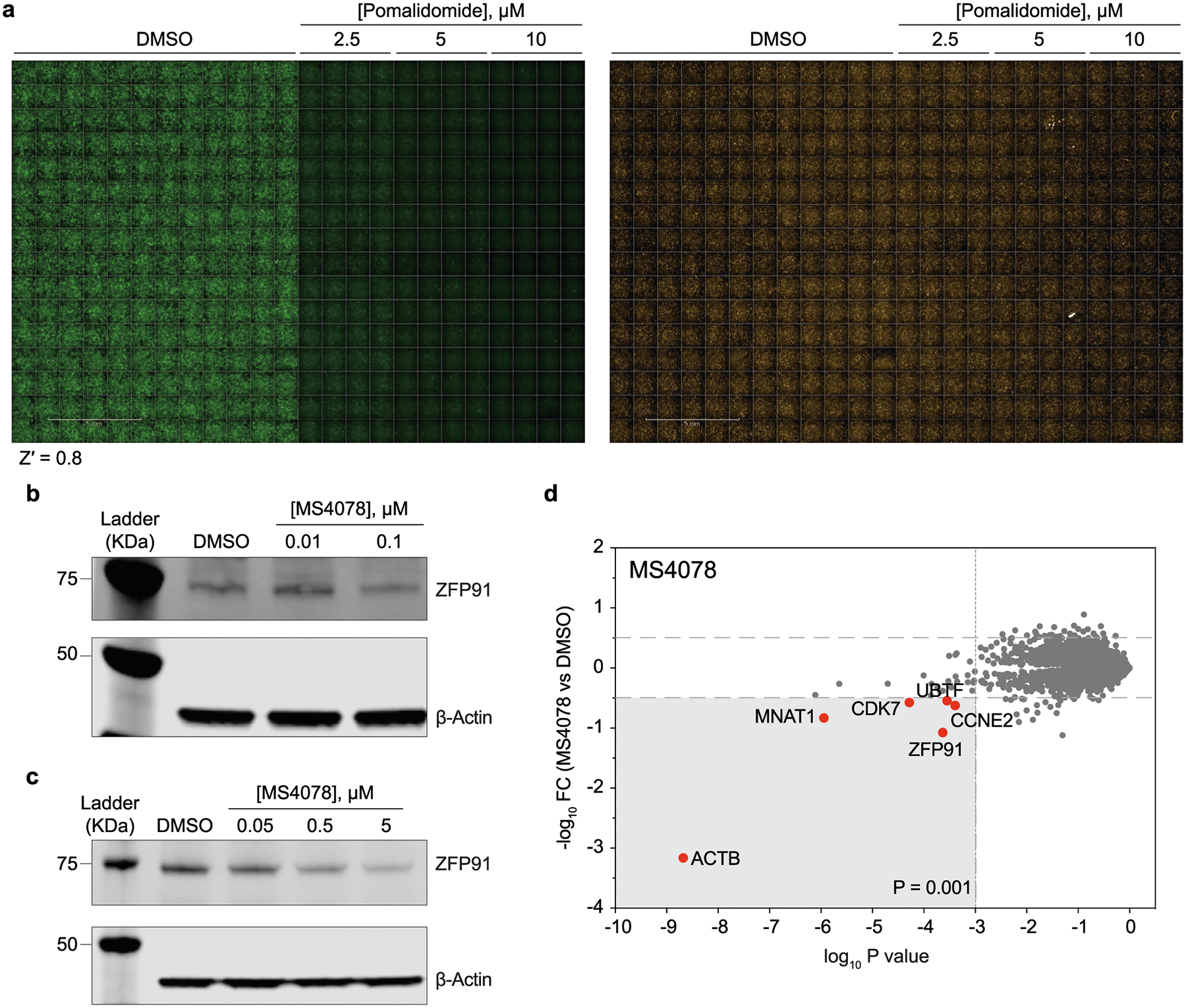
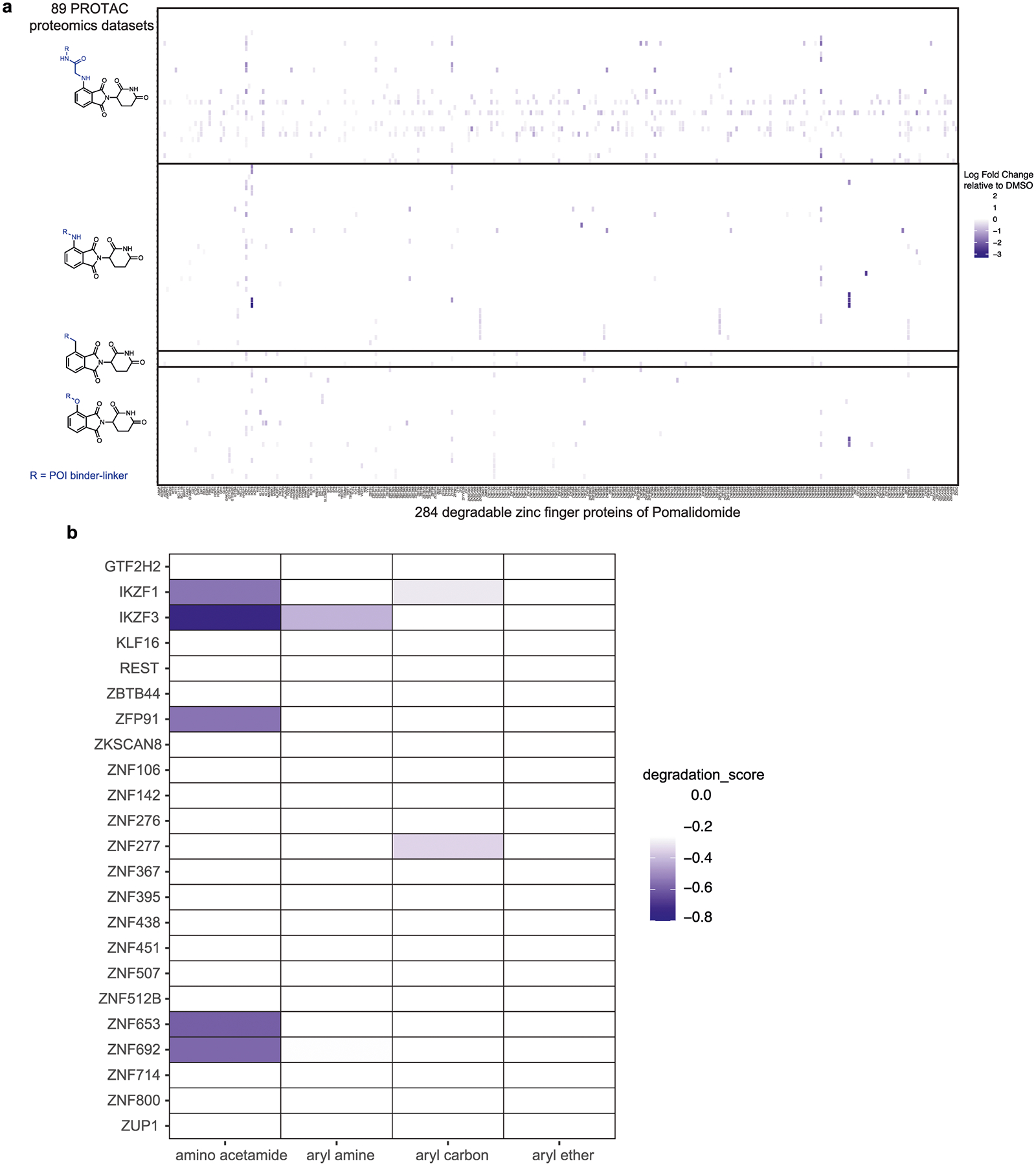
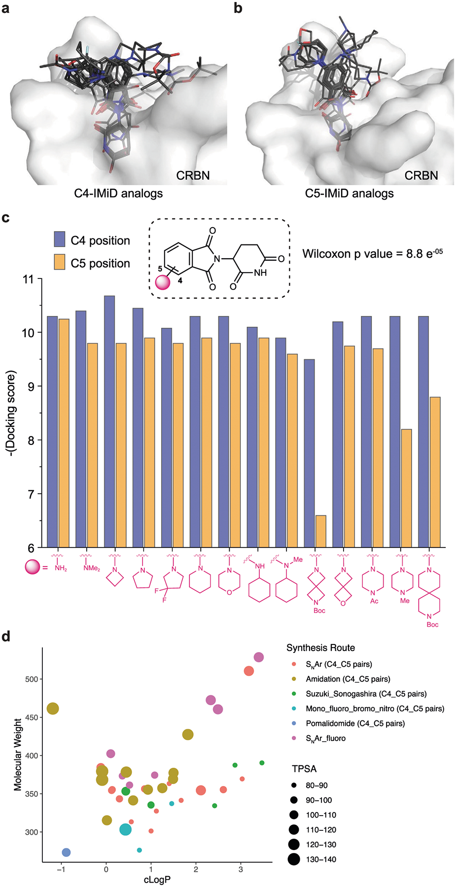
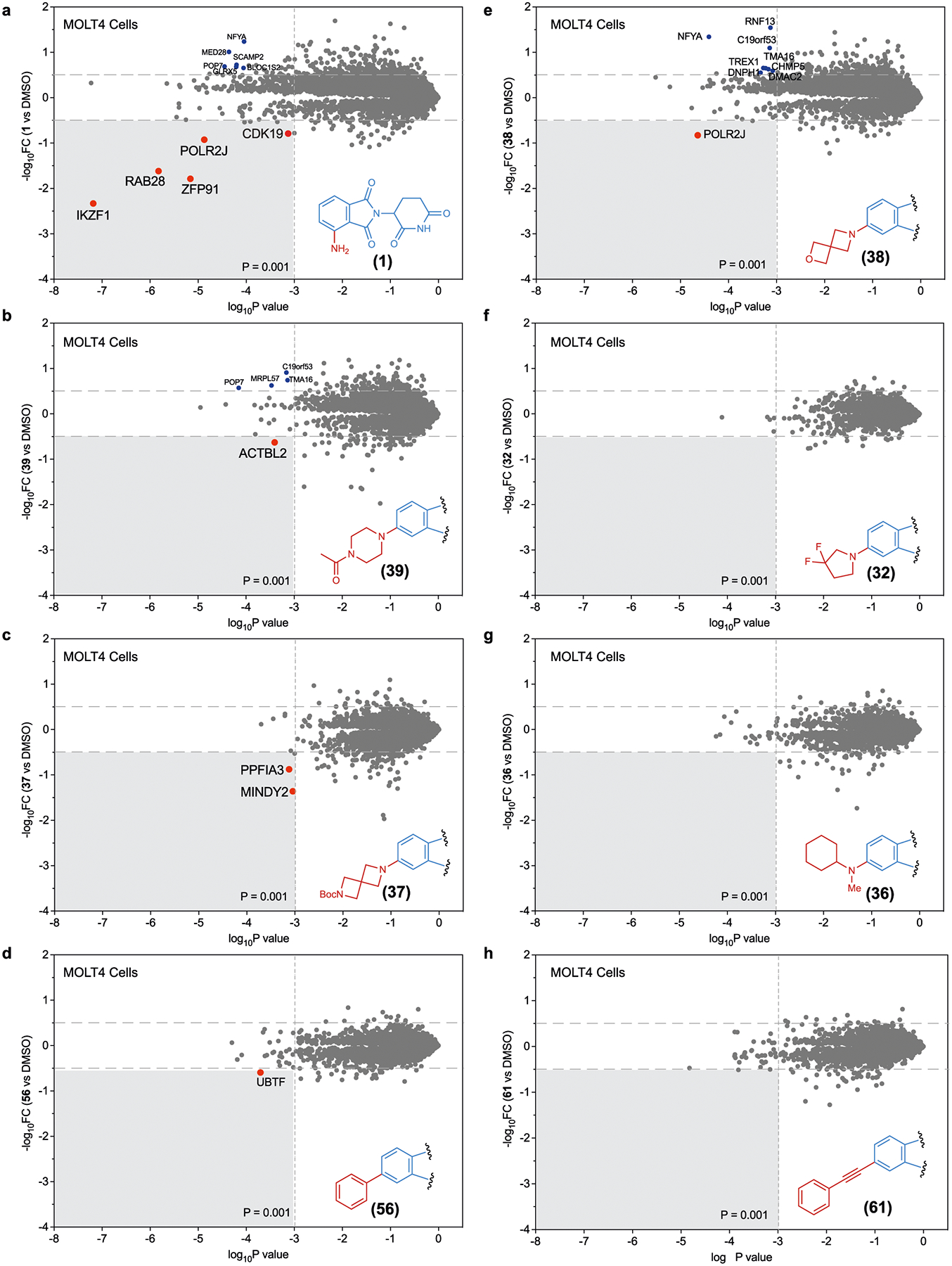
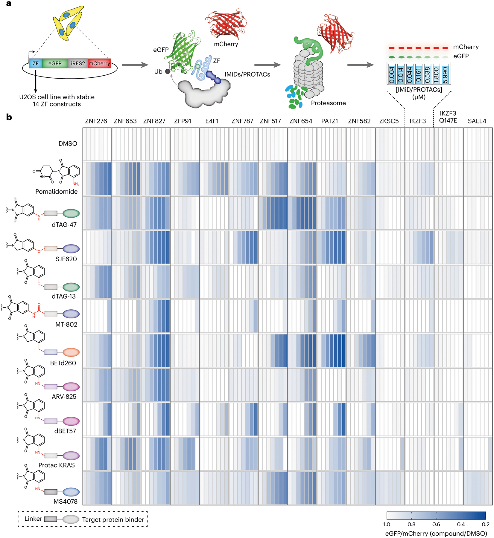
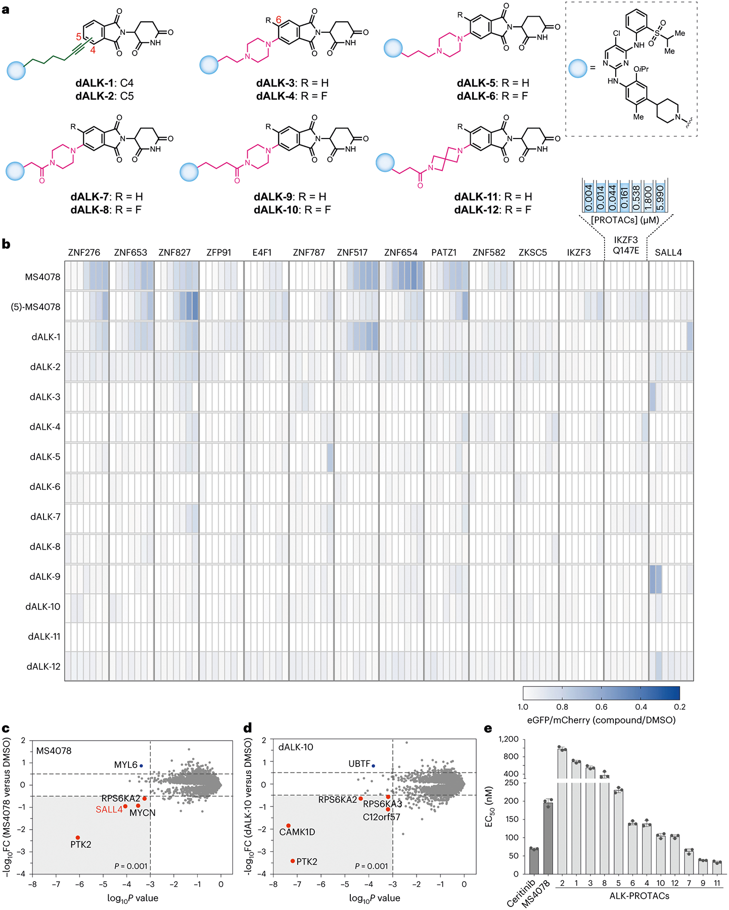
Proteolysis-targeting chimeras (PROTACs) are molecules that induce proximity between target proteins and E3 ligases triggering target protein degradation. Pomalidomide, a widely used E3 ligase recruiter in PROTACs, can independently degrade other proteins, including zinc-finger (ZF) proteins, with vital roles in health and disease. This off-target degradation hampers the therapeutic applicability of pomalidomide-based PROTACs, requiring development of PROTAC design rules that minimize off-target degradation. Here we developed a high-throughput platform that interrogates off-target degradation and found that reported pomalidomide-based PROTACs induce degradation of several ZF proteins. We generated a library of pomalidomide analogues to understand how functionalizing different positions of the phthalimide ring, hydrogen bonding, and steric and hydrophobic effects impact ZF protein degradation. Modifications of appropriate size on the C5 position reduced off-target ZF degradation, which we validated through target engagement and proteomics studies. By applying these design principles, we developed anaplastic lymphoma kinase oncoprotein-targeting PROTACs with enhanced potency and minimal off-target degradation.


Conflict of interest statement

Broad Institute has filed a patent application including the work described herein. A.C. is a founder and Scientific Advisory Board (SAB) member in Photys therapeutics. E.S.F. is a founder, SAB member, and equity holder in Civetta, Lighthorse, Proximity and Neomorph (board of directors), SAB member and equity holder in Photys and Avilar, and a consultant to Astellas, Novartis, Sanofi, Deerfield and EcoR1. The Fischer lab receives or has received research funding from Novartis, Astellas, Interline and Deerfield. D.R.L. is a consultant and co-founder of Exo Therapeutics, a company that develops small-molecule therapeutics. K.A.D. is a consultant to Kronos Bio and Neomorph Inc. The remaining authors declare no competing financial interests.

Figures

Extended Data Fig. 1 |
Extended Data Fig. 1 |. Establishment and validation of automated imaging assay for the degradation of ZFs.
a) Representative readouts of the imaging assay in 384-well plates, demonstrating robust detection of ZF-tagged eGFP degradation as induced by pomalidomide. Shown are images of U2OS cells with stable expression of pomalidomide-sensitive ZFP91 degron reporter. Z′ value was calculated using an in-built module in the Harmony software (See materials and methods). b, c) Immunoblots (from at least two independent replicates) quantifying off-target degradation of endogenous ZF proteins ZFP91 by MS4078 (ALK PROTAC) in a dose-dependent manner across cell lines SU-DHL-1 (b) and H2228 (c). d) Label-free proteomic analysis of MS4078 in MOLT4 cells.
Extended Data Fig. 2 |
Extended Data Fig. 2 |. Off-target ZF degradation assessed by mass spectrometry-based proteomics.
Identification of the same group of exit vectors on pomalidomide with minimal off-target ZF degradation assessed by mass spectrometry-based proteomics for entire data set (a) and top 10 most degraded proteins (b). Relative abundance of endogenous ZF proteins in cells treated with pomalidomide-based PROTACs as arranged based on pomalidomide’s exit vector groups. Data were extracted from proteomics datasets published in Donovan et al..
Extended Data Fig. 3 |
Extended Data Fig. 3 |. Docking scores and topological polar surface area of C4 and C5 pomalidomide analogs.
ac) Structural docking of pomalidomide analogs with C4 (a) and C5 (b) modifications on the phthalimide ring. Docking score (c) of each pair of modifications on C4 and C5 (paired Wilcoxon test, p = 8.8 × e-05). (d) Distribution of the physicochemical properties of the pomalidomide analog library. The topological polar surface area (TPSA) of each molecule is indicated by color and size (see legend). Note that each dot in the scatter plot represents a pair of compounds with the same modification on C4 and C5 positions, except the SNAr fluoro group. Synthetic routes are represented by different shapes shown in the legend).
Extended Data Fig. 4 |
Extended Data Fig. 4 |. Degradation of validated pomalidomide-sensitive ZF degrons induced by the pomalidomide analogs.
(a) Normalized eGFP intensity in 14 ZF reporter cell lines treated with different doses of 81 pomalidomide analogs ranging from 4.3 nM to 20 μM. Each block of 4.3 nM to 20 μM doses on the x axis represents one ZF reporter cell line. Data are mean of at least 3 independent replicates. (See source data for replicates, mean, SD) (bd) Box-and-Whisker plots with statistical analysis for pomalidomide analogs arranged in pairs of C4 and C5 modifications such as acylation (b), Suzuki/Sonogashira coupling (c) and effect of -F group (d) on the phthalimide ring. Data are shown for cells treated with 5 μM of each compound (for acylated and Suzuki/Sonogashira couplings) and 20 μM data of each compound for deciphering the -F group effect. For b-d figure panels data points are the mean of at least 3 independent replicates. Centre of the box plot is median. Minima and maxima are 1.5 times the interquartile range over the 75th and under the 25th percentile, respectively.
Extended Data Fig. 5 |
Structures of IMiD analogs with the least degradation score (close to 0).
Extended Data Fig. 6 |
NanoBRET based ternary complex analysis of all the IMiD analogs and PROTACs reported in this study.
Extended Data Fig. 7 |
Global Proteomic analysis in KELLY cells. a–h, Global proteomic analysis of Kelly cells treated with analogues 1 (a), 39 (b), 37 (c), 56 (d), 38 (e), 32 (f), 36 (g) and 61 (h). FC, fold change.
Extended Data Fig. 8 |
Extended Data Fig. 8 |. Cellular characterization of dALK PROTACs.
a, b) Global Proteomic analysis of selected PROTACs, MS4078 (a) and dALK-10 (b) in SU-DHL-1 cells. c) Viability dose curves for the redesigned ALK PROTACs in SU-DHL-1 cells. Data are the mean ± SD of 3 independent replicates.
Fig. 1 |
Fig. 1 |. Development of a high-throughput assay for evaluating off-target ZF degradation of pomalidomide-based PROTACs.
a, An automated imaging screen for the degradation of ZF degrons by pomalidomide analogues and pomalidomide-based PROTACs. Briefly, U2OS cells stably expressing 14 ZF degrons fused to eGFP were treated with PROTACs followed by imaging to assess ZF degradation. Decrease in the eGFP signal is the measure of ZF protein degradation. b, Degradation of validated and pomalidomide-sensitive ZF degrons inside cells by reported PROTACs in a dosing range of 4.3 nM to 6 μM. As shown in the schematic, each rectangle in the heatmap represents a single dose. Chemical structures with rectangle boxes and oval shapes indicate linker chemotype and target protein binding ligands respectively. Data are the mean of at least three independent replicates.
Fig. 2 |
Fig. 2 |. Validation of image-based results using NanoBRET and immunoblotting.
Structural insights and degradation potential of the thalidomide analogues with C4 and C5 amino groups on the phthalimide ring. a, A NanoBRET assay to quantify ternary complex formation between ZFP91, CRBN and imide analogues or PROTACs. b, BRET ratio of reported PROTACs treated at 1 μM dose in a NanoBRET assay. Data are the mean of two independent replicates. c,d, Representative immunoblots (from at least two independent replicates) demonstrating off-target degradation of endogenous ZF protein ZFP91 by MS4078 (ALK PROTAC) (c) and dTAG-13 (FKBP12F36V PROTAC) (d) in a dose-dependent manner in Jurkat cells. e, Crystal structure showing the glutarimide ring deeply buried in the CRBN exposing the 4-amino group of pomalidomide, which makes a crucial water-mediated hydrogen bond between CRBN (E377) and IKZF1 (Q147). Modification on C5 position would potentially bump off the ZF degrons (PDB: 6H0F). f, Structures of pomalidomide (1) and 5-aminothalidomide (8). g, Representative immunoblots (from at least two independent replicates) of endogenous ZF proteins ZFP91 and IKZF3 in MM1.S cells treated with pairs of imide-based analogues with C4 (pomalidomide) and C5 amino modifications on the phthalimide ring.
Fig. 3 |
Fig. 3 |
Synthesis of imide analogues via nucleophic aromatic substitution (SNAr) reactions.
Fig. 4 |
Fig. 4 |. Synthesis of imide analogs using bromo- and amino- imide building blocks.
a,b, Synthesis of imide analogues via cross-coupling (a) and amidation (b) chemistries.
Fig. 5 |
Fig. 5 |. Degradation score as metric to nominate the imide analogues with reduced off-targets and cellular target engagement studies.
a, Plot showing pair-wise comparison of GFP degradation levels induced by pairs of SNAr pomalidomide analogues with C4 and C5 modifications (Wilcoxon matchedpairs signed-rank test; P = 0.0029) at the same dose. Data are the mean of at least three independent replicates. Centre of the box plot is median. Minima and maxima are 1.5 times the interquartile range over the 75th and under the 25th percentile, respectively. b, Degradation of pomalidomide-sensitive ZF degrons inside cells by pomalidomide analogues with and without H-bond donor(s) immediately adjacent to the phthalimide ring. Data are the mean of at least three independent replicates. (two tailed Welch’s t-test; P < 0.0001). Centre of the box plot is median. Minima and maxima are 1.5 times the interquartile range over the 75th and under the 25th percentile, respectively. c, Representative immunoblots (from at least two independent replicates) for endogenous ZF proteins ZFP91 and IKZF3 in Jurkat cells treated with pomalidomide analogues (21 and 22) with and without a H-bond donor (NH) immediately attached to the phthalimide ring. d, Scatter dot plot showing ZF degradation scores (that is, −log(degradation sum + 1)) for individual pomalidomide analogues and pomalidomide-based PROTACs investigated in this study. Data are the mean of at least three independent replicates. (C5 -F indicate fluoro substitution on C5) e, A competition-based NanoBRET assay for CRBN engagement in cells by the pomalidomide analogues. f, Dose-dependent CRBN binding curves of selected analogues determined in U2OS cells using NanoBRET-based intracellular CRBN binding assay. Data are the mean of at least four independent replicates. g, BRET ratio for selected analogues treated at 1 μM dose in a NanoBRET based ternary complex formation assay (for the ternary complex analysis of all the imide analogues generated in this study, see Supplementary Fig. 9). Data are the mean two independent replicates.
Fig. 6 |
Fig. 6 |. Validation of image-based platform by global proteomics.
ah, Global proteomic analysis of MOLT4 cells treated with analogues 1 (a), 39 (b), 37 (c), 56 (d), 38 (e), 32 (f), 36 (g) and 61 (h). FC, fold change.
Fig. 7 |
Fig. 7 |. Re-engineering of ALK PROTACs based on the design principles.
Validation of image-based degradation studies. a, Structures of rationally redesigned ALK PROTACs to minimize off-target ZF degradation. b, Degradation of validated and pomalidomide-sensitive ZF degrons inside cells by redesigned ALK PROTACs in a dosing range of 4.3 nM to 6 μM. Data are the mean of three independent replicates. c,d, Global proteomic analysis of KELLY cells treated with 100 nM of MS4078 (c) or dALK-10 (d), respectively. e, Determination of EC50 values (nM) of SU-DH-L1 cells upon treatment with redesigned ALK PROTACs. Error bars represent mean ± standard deviation from three biological replicates. FC, fold change.

References

    1. Chamberlain PP & Cathers BE Cereblon modulators: low molecular weight inducers of protein degradation. Drug Discov. Today Technol 31, 29–34 (2019). - PubMed
    1. Kozicka Z & Thomä NH Haven’t got a glue: protein surface variation for the design of molecular glue degraders. Cell Chem. Biol 28, 1032–1047 (2021). - PubMed
    1. Finley D Recognition and processing of ubiquitin–protein conjugates by the proteasome. Annu. Rev. Biochem 78, 477–513 (2009). - PMC - PubMed
    1. Ito T et al. Identification of a primary target of thalidomide teratogenicity. Science 327, 1345–1350 (2010). - PubMed
    1. Krönke J et al. Lenalidomide causes selective degradation of IKZF1 and IKZF3 in multiple myeloma cells. Science 343, 301–305 (2014). - PMC - PubMed

MeSH terms