. 2023 Dec 19;13(1):22562.
doi: 10.1038/s41598-023-49881-w.

S9.6-based hybrid capture immunoassay for pathogen detection

Affiliations

S9.6-based hybrid capture immunoassay for pathogen detection

Ankur Bothra et al. Sci Rep. .

Abstract

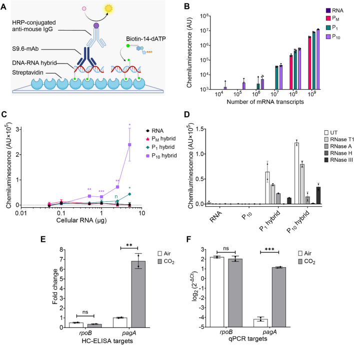
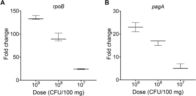
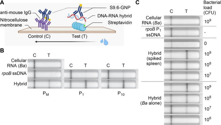
The detection of pathogens is critical for clinical diagnosis and public health surveillance. Detection is usually done with nucleic acid-based tests (NATs) and rapid antigen tests (e.g., lateral flow assays [LFAs]). Although NATs are more sensitive and specific, their use is often limited in resource-poor settings due to specialized requirements. To address this limitation, we developed a rapid DNA-RNA Hybrid Capture immunoassay (HC) that specifically detects RNA from pathogens. This assay utilizes a unique monoclonal antibody, S9.6, which binds DNA-RNA hybrids. Biotinylated single-stranded DNA probes are hybridized to target RNAs, followed by hybrid capture on streptavidin and detection with S9.6. The HC-ELISA assay can detect as few as 104 RNA molecules that are 2.2 kb in length. We also adapted this assay into a LFA format, where captured Bacillus anthracis rpoB RNA of 3.5 kb length was detectable from a bacterial load equivalent to 107 CFU per 100 mg of mouse tissue using either HC-ELISA or HC-LFA. Importantly, we also demonstrated the versatility of HC by detecting other pathogens, including SARS-CoV-2 and Toxoplasma gondii, showing its potential for broad pathogen detection. Notably, HC does not require amplification of the target nucleic acid and utilizes economical formats like ELISA and LFA, making it suitable for use in sentinel labs for pathogen detection or as a molecular tool in basic research laboratories. Our study highlights the potential of HC as a sensitive and versatile method for RNA-based pathogen detection.


Conflict of interest statement

The authors declare no competing interests.

Figures

Figure 1
Figure 1
Analysis of S9.6’s specificity for DNA-RNA hybrids. (A) Representative dot blot analysis of S9.6 binding to a crude extract of cellular RNA from B. anthracis. One microgram of cellular RNA was spotted directly on a nitrocellulose membrane or first treated with respective nucleases. UT untreated, MBN mung bean nuclease. Nuclease specificities are as follows: RNase H, DNA-RNA hybrids; MBN, ssDNA; RNase T1, ssRNA; RNase III, dsRNA; RNase A, all RNA. (B) Quantification of signal intensity relative to untreated (UT) control for nuclease treated samples. Data represents mean ± SD of duplicate dot blots (A, Fig. S1A). (C) Dot blot analysis using S9.6 to detect in vitro synthesized pagA mRNA, ssDNA, and DNA-RNA hybrids in specified amounts.
Figure 2
Figure 2
Sensitivity and specificity of HC-ELISA to detect B. anthracis gene transcripts. (A) Schematic of the HC-ELISA format. Gene specific DNA-RNA hybrids are generated using 5’-biotinylated ssDNA probes (PM) or 5′-biotinylated ssDNA probes synthesized in the presence of 1 µM (P1) or 10 µM (P10) biotin-14-dATP. The hybrids are captured on a poly-streptavidin coated ELISA plate and bound to S9.6, which is detected using anti-mouse IgG conjugated with HRP (horseradish peroxidase) and a chemiluminescent substrate. Illustration created with BioRender.com. (B) HC-ELISA sensitivity to pagA hybrids incorporating PM, P1, and P10 probes. Data represents mean arbitrary units (AU) of chemiluminescence normalized to average of ssDNA and buffer-alone control ± SD (n = 3 wells). (C) Signal from HC-ELISAs detecting rpoB transcripts in B. anthracis cellular RNA. Data represent mean chemiluminescence normalized to average of ssDNA and buffer-alone control ± SD (n = 2 wells). (D) Assay specificity assessment using ribonucleases. Data represent mean chemiluminescence normalized to average of ssDNA and buffer-alone control ± SD (n = 2 wells). UT = untreated. (E) rpoB and pagA expression over reference gene gyrA between ambient and CO2-rich conditions as measured by chemiluminescence from HC-ELISA and (F) relative abundance of rpoB and pagA to gyrA as measured by RT-qPCR. Error bars represent mean ± SD of duplicates; statistical significance was determined with Student’s t-test. *p < 0.05, **p < 0.01, ***p < 0.001; ns, not significant. Panels (BD) show a representative experiment of three repeated experiments; panels (E,F) present the data in full.
Figure 3
Figure 3
HC-ELISA can detect simulated B. anthracis infection in mice. (A,B) Median fold change in chemiluminescence as a function of bacterial CFU/100 mg of spleen suspension as measured by HC-ELISAs detecting rpoB (A) and pagA (B). Fold change was calculated relative to uninfected control spleens. Error bars represent range of triplicate wells in a representative experiment of n = 3.
Figure 4
Figure 4
HC-LFA can detect simulated B. anthracis infections in mice. (A) Schematic of HC-LFA. S9.6-GNP = gold nanoparticle-conjugated S9.6. The arrow indicates the direction of mobile phase. Illustration created with BioRender.com. (B) Images of lateral flow assays showing HC-LFA’s specificity for hybrids by probe type (B. anthracis RNA, 2 µg). (C) Images of lateral flow assays showing HC-LFA to detect rpoB transcripts in cellular RNA from B. anthracis or B. anthracis-spiked murine spleen tissue. The spiked bacterial load ranged from 107 to 109 CFU/100 mg of tissue. For bacteria alone, 2 µg cellular RNA, equivalent to a bacterial load of 109 CFU was diluted. 5 ng of P1 probe was used in hybridization and ssDNA controls. Assays were imaged after 30 min. Individual lateral flow strips are placed separately and are grouped within the dashed line in (B) and (C).
Figure 5
Figure 5
HC assays can detect RNA transcripts from T. gondii and SARS-CoV-2. (A) Average fold change in chemiluminescence from HC-ELISA to detect B1 gene mRNA of T. gondii-infected HFF cells over uninfected cells seeded at the same density at start of study and harvested at the same time as infected cells. The P10 probe was used in this experiment and C (control) is the probe hybridized with cellular RNA from overly confluent uninfected cells. Bars represent mean ± SD of duplicate wells in a representative experiment of n = 3. (B) Median chemiluminescence from HC-ELISA to detect in vitro synthesized Spike E mRNA of SARS-CoV-2. Data obtained using 2 × 109 molecules/well of specified nucleic acids (assuming full hybridization from equal amounts RNA and probe for P10 hybrid). Bars represent range of triplicate wells in a representative experiment of n = 3. (C) Lateral flow assays show the sensitivity of HC-LFA to Spike E gene hybrids generated from 5 ng (2.6 × 109 molecules) P1 probe and in vitro-synthesized mRNA in amounts specified. Assays were imaged after 30 min. Individual lateral flow strips are placed separately and are grouped within the dashed line.

Similar articles

Cited by

References

    1. Emmadi R, et al. Molecular methods and platforms for infectious diseases testing a review of FDA-approved and cleared assays. J. Mol. Diagn. 2011;13:583–604. doi: 10.1016/j.jmoldx.2011.05.011. - DOI - PMC - PubMed
    1. Lau HY, Botella JR. Advanced DNA-based point-of-care diagnostic methods for plant diseases detection. Front. Plant Sci. 2017;8:2016. doi: 10.3389/fpls.2017.02016. - DOI - PMC - PubMed
    1. Valkiunas G, et al. A comparative analysis of microscopy and PCR-based detection methods for blood parasites. J. Parasitol. 2008;94:1395–1401. doi: 10.1645/GE-1570.1. - DOI - PubMed
    1. Pilotte N, et al. Improved PCR-based detection of soil transmitted helminth infections using a next-generation sequencing approach to assay design. PLoS Negl. Trop. Dis. 2016;10:e0004578. doi: 10.1371/journal.pntd.0004578. - DOI - PMC - PubMed
    1. Fozouni, P. et al. Amplification-free detection of SARS-CoV-2 with CRISPR-Cas13a and mobile phone microscopy. Cell184, 323–333 e329 (2021). - PMC - PubMed