. 2023 Dec 18;3(1):186.
doi: 10.1038/s43856-023-00381-y.

Concurrent gliomas in patients with multiple sclerosis

Affiliations

Concurrent gliomas in patients with multiple sclerosis

Katharina Sahm et al. Commun Med (Lond). .

Abstract

Background: Concurrent malignant brain tumors in patients with multiple sclerosis (MS) constitute a rare but paradigmatic phenomenon for studying neuroimmunological mechanisms from both molecular and clinical perspectives.

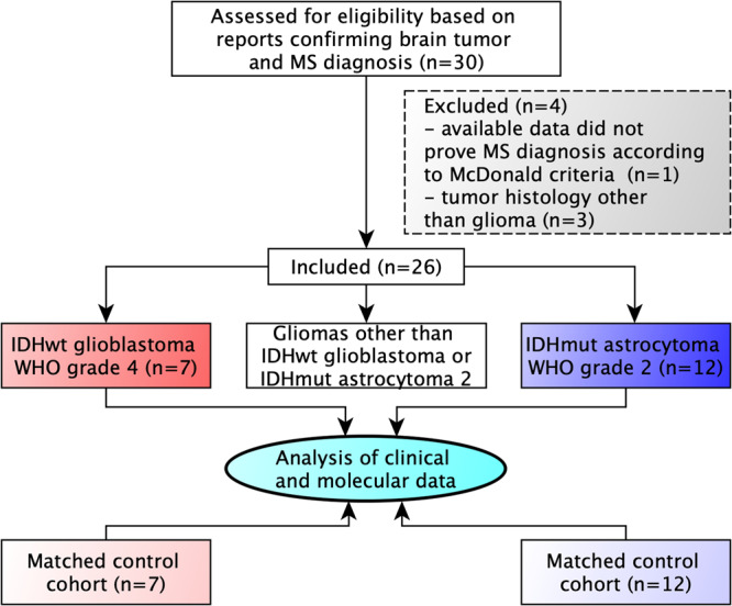
Methods: A multicenter cohort of 26 patients diagnosed with both primary brain tumors and multiple sclerosis was studied for disease localization, tumor treatment-related MS activity, and molecular characteristics specific for diffuse glioma in MS patients.

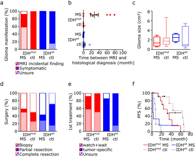
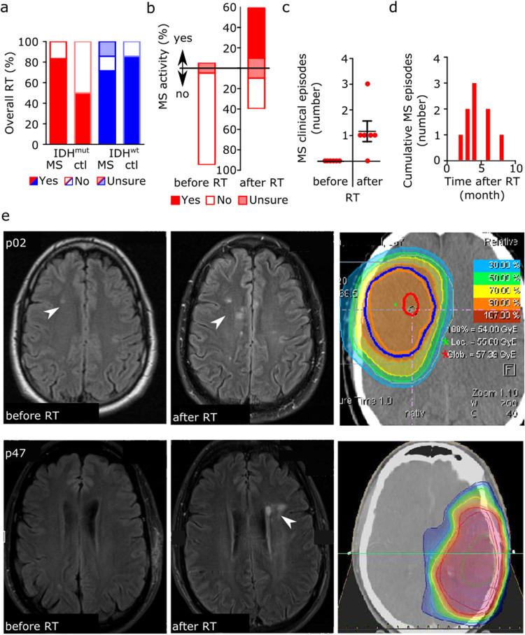
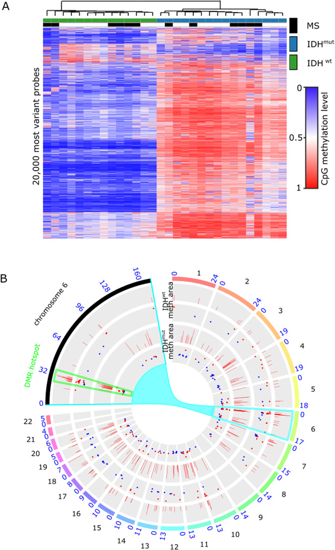
Results: MS neither predisposes nor protects from the development of gliomas. Patients with glioblastoma WHO grade 4 without isocitratdehydrogenase (IDH) mutations have a longstanding history of MS, whereas patients diagnosed with IDH-mutant astrocytoma WHO grade 2 receive multiple sclerosis diagnosis mostly at the same time or later. Concurrent MS is associated with a lesser extent of tumor resection and a worse prognosis in IDH-mutant glioma patients (PFS 32 vs. 64 months, p = 0.0206). When assessing tumor-intrinsic differences no distinct subgroup-defining methylation pattern is identified in gliomas of MS patients compared to other glioma samples. However, differential methylation of immune-related genetic loci including human leukocyte antigen locus on 6p21 and interleukin locus on 5q31 is found in MS patients vs. matched non-MS patients. In line, inflammatory disease activity increases in 42% of multiple sclerosis patients after brain tumor radiotherapy suggesting a susceptibility of multiple sclerosis brain tissue to pro-inflammatory stimuli such as ionizing radiation.

Conclusions: Concurrent low-grade gliomas should be considered in multiple sclerosis patients with slowly progressive, expansive T2/FLAIR lesions. Our findings of typically reduced extent of resection in MS patients and increased MS activity after radiation may inform future treatment decisions.

Plain language summary

Brain tumors such as gliomas can evade attacks by the immune system. In contrast, some diseases of the central nervous system such as multiple sclerosis (MS) are caused by an overactive immune system. Our study looks at a cohort of rare patients with both malignant glioma and concurrent MS and examines how each disease and their treatments affect each other. Our data suggest that even in patients with known MS, if medical imaging findings are unusual, a concurrent brain tumor should be excluded at an early stage. Radiotherapy, as is the standard of care for malignant brain tumors, may worsen the inflammatory disease activity in MS patients, which may be associated with certain genetic risk factors. Our findings may help to inform treatment of patients with brain tumors and MS.


Conflict of interest statement

The authors declare no competing interests.

Figures

Fig. 1
Fig. 1. Flow diagram of the multicenter study.
Diagram showing the number and main inclusion and exclusion criteria of recruited patients as well as group allocation.
Fig. 2
Fig. 2. Clinical and radiological characteristics of 19 patients with multiple sclerosis and concurrent glioma.
a, b Scatter dot plot of patients’ age at initial histologically proven diagnosis of glioma depending on the mutation status of isocitrate dehydrogenase-1 (IDH) in patients with concurrent multiple sclerosis (MS) or control patients without multiple sclerosis (ctl) (a), and patients’ age at initial multiple sclerosis diagnosis depending on tumoral IDH mutation status (b). Depicted are single and mean values with SEM (IDH-mut MS n = 12; IDH-wt MS n = 7). c Chronology of multiple sclerosis and glioma diagnosis depending on tumor histology judged by IDH mutation status. d Schematic representation of glioma location in patients with concurrent multiple sclerosis depending on tumoral IDH mutation status. Dots represent single patients. e MRI with axial FLAIR and contrast-enhanced T1 images of a patient with multiple sclerosis and concurrent IDH-mutant astrocytoma WHO grade 2 (p02, upper row) and of a patient with multiple sclerosis and IDH-wildtype glioblastoma WHO grade 4 (p08, lower row). f Schematic representation of the spatial relation of multiple sclerosis lesions and IDH-mutant astrocytomas (red) or IDH-wildtype glioblastomas (blue) in patients with concurrent diseases.
Fig. 3
Fig. 3. Clinical course of brain tumor disease in patients with glioma and concurrent multiple sclerosis.
a Percentage of patients with or without glioma-caused neurological symptoms leading to initial tumor diagnosis in patients with concurrent multiple sclerosis (MS) or matched controls (ctl) depending on tumoral isocitrate dehydrogenase-1 (IDH) mutation status. b Time between radiological tumor suspicion and subsequent histological diagnosis. Depicted are single and mean values with SEM (IDH-mut MS n = 10; IDH-mut ctl n = 12; IDH-wt MS n = 6; IDH-wt ctl n = 7). c Tumor size as detected by MRI at the time of histological diagnosis in patients with concurrent multiple sclerosis or matched controls depending on tumoral IDH mutation status. Displayed are the products of the two largest perpendicular diameters (cm2) in axial FLAIR or contrast-enhanced T1 images for IDH-mutant astrocytomas WHO grade 2 and IDH-wildtype glioblastomas, respectively. Depicted are mean values with SEM and min to max (IDH-mut MS n = 8; IDH-mut ctl n = 8; IDH-wt MS n = 2; IDH-wt ctl n = 7). d Percentage of patients receiving tumor biopsy, partial, or complete resection. e Type of adjuvant tumor therapy depending on concurrent multiple sclerosis diagnosis and IDH mutation status. f Kaplan–Meier analysis of progression-free survival (PFS) after primary tumor treatment depending on IDH status and concurrent multiple sclerosis diagnosis (IDH-mut MS n = 12; IDH-mut ctl n = 12; IDH-wt MS n = 7; IDH-wt ctl n = 7).
Fig. 4
Fig. 4. Association between radiotherapy and multiple sclerosis disease activity.
a Percentage of patients with or without radiotherapy (RT) treatment during the course of brain tumor disease depending on concurrent multiple sclerosis (MS) diagnosis and tumoral IDH mutation status. b Percentage of patients with or without clinical or radiological MS disease activity in the 12 months preceding (left bar) and after RT (right bar). c Scatter dot plot of the number of clinical episodes per patient for six multiple sclerosis patients in the 12 months preceding and after RT. Depicted are single and mean values with SEM. d Time course of cumulative clinical multiple sclerosis episodes for six multiple sclerosis patients experiencing disease activity in the 12 months after RT. Individual number of clinical episodes per patient are one episode in 4/6 patients, two episodes in 1/6 patients and three episodes in 1/6 patients. e Magnet resonance imaging (MRI) with axial FLAIR images of two patients with isocitrate dehydrogenase-1 (IDH)-mutant astrocytomas showing new demyelinating lesions (arrows) after RT within the 50% isodose line (p02 photon irradiation 60/2 Gy; p47 proton irradiation 54/1.8 Gy).
Fig. 5
Fig. 5. Long-term response after radiation-induced pseudoprogression.
Representation of isodoses (Gy) and longitudinal MRIs with axial FLAIR images (A) and contrast-enhanced axial T1 images (B) of a patient (p08) with isocitrate dehydrogenase-1 (IDH)-wildtype glioblastoma WHO grade 4. Transient contrast enhancement within the tumor area corresponding to radiation-induced pseudoprogression with a maximum of 8 months after completion of radiotherapy (60/2 Gy) is associated with an increase in subcortical multiple sclerosis lesions (arrows) and the initial diagnosis of a chronic progressive multiple sclerosis course. Long term follow-up shows a moderate generalized brain atrophy (stars) without evidence of glioblastoma recurrence up to 8 years after initial tumor therapy.
Fig. 6
Fig. 6. Genome-wide DNA methylation changes between gliomas of patients with concurrent multiple sclerosis and controls.
A Unsupervised hierarchical clustering of methylation profiles from 450k methylation array data of 12 glioma tissue in multiple sclerosis (MS) patients and matched controls. B Circos plot showing differentially methylated regions (DMR) between patients with and without multiple sclerosis separately for isocitrate dehydrogenase-1-mutant (IDHmut, inner two circles) and isocitrate dehydrogenase-1-wildtype tumors (IDHwt, outer two circles) on each of the human chromosomes with a zoom in on chromosome 6. The inner circle of the respective comparisons show the average methylation, the outer circle the area of the DMR.

References

    1. Sinclair G, Al-Saffar Y, Johnstone P, Hatiboglu MA, Shamikh A. A challenging case of concurrent multiple sclerosis and anaplastic astrocytoma. Surgical Neurol. Int. 2019;10:166–166. doi: 10.25259/SNI_176_2019. - DOI - PMC - PubMed
    1. Preziosa P, et al. Clinical deterioration due to co-occurrence of multiple sclerosis and glioblastoma: report of two cases. Neurol. Sci. 2017;38:361–364. doi: 10.1007/s10072-016-2763-y. - DOI - PubMed
    1. Plantone D, Renna R, Sbardella E, Koudriavtseva T. Concurrence of multiple sclerosis and brain tumors. Front. Neurol. 2015;6:40. doi: 10.3389/fneur.2015.00040. - DOI - PMC - PubMed
    1. Acqui M, Caroli E, Di Stefano D, Ferrante L. Cerebral ependymoma in a patient with multiple sclerosis case report and critical review of the literature. Surg. Neurol. 2008;70:414–420. doi: 10.1016/j.surneu.2007.04.015. - DOI - PubMed
    1. Sierra Morales F, et al. Glioblastoma in natalizumab-treated multiple sclerosis patients. Ann. Clin. Transl. Neurol. 2017;4:512–516. doi: 10.1002/acn3.428. - DOI - PMC - PubMed