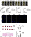
PAK inhibitor FRAX486 decreases the metastatic potential of triple-negative breast cancer cells by blocking autophagy
- PMID: 38110664
- PMCID: PMC10844298
- DOI: 10.1038/s41416-023-02523-4
PAK inhibitor FRAX486 decreases the metastatic potential of triple-negative breast cancer cells by blocking autophagy
Abstract
Background: Triple-negative breast cancer (TNBC) is a unique breast cancer subtype with a high risk of metastasis and recurrence and a poor prognosis. Epithelial-mesenchymal transition (EMT) endows epithelial cells with the ability to move to distant sites, which is essential for the metastasis of TNBC to organs, including the lung. Autophagy, an intracellular degradation process that involves formation of double-layered lipid autophagosomes that transport cytosolic cargoes into lysosomes via autophagosome-lysosome fusion, is involved in various diseases, including cancer and neurodegenerative, metabolic, cardiovascular, and infectious diseases. The relationship between autophagy and cancer has become relatively clear. However, research on pharmacological drugs that block cancer EMT by targeting autophagy is still in the initial stages. Therefore, the re-evaluation of old drugs for their potential in blocking both autophagy and EMT was conducted.
Methods: More than 2000 small molecule chemicals were screened for dual autophagy/EMT inhibitors, and FRAX486 was identified as the best candidate inhibitor of autophagy/EMT. The functions of FRAX486 in TNBC metastasis were detected by CCK-8, migration and wound healing assays. The effects of FRAX486 on autophagy and its target PAK2 were determined by immunoblotting, immunofluorescence, immunoprecipitation analysis and transmission electron microscopy. The findings were validated in mouse models.
Results: Here, we report that FRAX486, a potent P21-activated kinase 2 (PAK2) inhibitor, facilitates TNBC suppression both in vitro and in vivo by blocking autophagy. Mechanistically, FRAX486 inhibits autophagy in TNBC cells by targeting PAK2, leading to the ubiquitination and proteasomal degradation of STX17, which mediates autophagosome-lysosome fusion. The inhibition of autophagy by FRAX486 causes upregulation of the epithelial marker protein E-cadherin and thus suppresses the migration and metastasis of TNBC cells.
Conclusions: The effects of FRAX486 on TNBC metastasis suppression were verified to be dependent on PAK2 and autophagy inhibition. Together, our results provide a molecular basis for the application of FRAX486 as a potential treatment for inhibiting the metastasis of TNBC.
© 2023. The Author(s).
Conflict of interest statement
The authors declare no competing interests.
Figures


Similar articles
-
Eribulin mesilate suppresses experimental metastasis of breast cancer cells by reversing phenotype from epithelial-mesenchymal transition (EMT) to mesenchymal-epithelial transition (MET) states.Br J Cancer. 2014 Mar 18;110(6):1497-505. doi: 10.1038/bjc.2014.80. Epub 2014 Feb 25. Br J Cancer. 2014. PMID: 24569463 Free PMC article.
-
PKMYT1 Promotes Epithelial-Mesenchymal Transition Process in Triple-Negative Breast Cancer by Activating Notch Signaling.Rev Invest Clin. 2024;76(1):45-59. doi: 10.24875/RIC.23000256. Rev Invest Clin. 2024. PMID: 38442372
-
MicroRNA-1246 suppresses the metastasis of breast cancer cells by targeting the DYRK1A/PGRN axis to prevent the epithelial-mesenchymal transition.Mol Biol Rep. 2022 Apr;49(4):2711-2721. doi: 10.1007/s11033-021-07080-8. Epub 2022 Jan 21. Mol Biol Rep. 2022. PMID: 35059968
-
DNMT1: A key drug target in triple-negative breast cancer.Semin Cancer Biol. 2021 Jul;72:198-213. doi: 10.1016/j.semcancer.2020.05.010. Epub 2020 May 24. Semin Cancer Biol. 2021. PMID: 32461152 Review.
-
The role of EMT-related lncRNA in the process of triple-negative breast cancer metastasis.Biosci Rep. 2021 Feb 26;41(2):BSR20203121. doi: 10.1042/BSR20203121. Biosci Rep. 2021. PMID: 33443534 Free PMC article. Review.
Cited by
-
The up-regulation of PAK2 indicates unfavorable prognosis in patients with serous epithelial ovarian cancer and contributes to paclitaxel resistance in ovarian cancer cells.BMC Cancer. 2024 Sep 30;24(1):1213. doi: 10.1186/s12885-024-12969-1. BMC Cancer. 2024. PMID: 39350056 Free PMC article.
-
Tetrahydropyrazolopyridinones as a Novel Class of Potent and Highly Selective LIMK Inhibitors.J Med Chem. 2025 Aug 28;68(16):17427-17456. doi: 10.1021/acs.jmedchem.5c00974. Epub 2025 Aug 6. J Med Chem. 2025. PMID: 40765414 Free PMC article.
-
The Role of the p21-Activated Kinase Family in Tumor Immunity.Int J Mol Sci. 2025 Apr 20;26(8):3885. doi: 10.3390/ijms26083885. Int J Mol Sci. 2025. PMID: 40332759 Free PMC article. Review.
-
Epithelial-mesenchymal plasticity in cancer: signaling pathways and therapeutic targets.MedComm (2020). 2024 Aug 1;5(8):e659. doi: 10.1002/mco2.659. eCollection 2024 Aug. MedComm (2020). 2024. PMID: 39092293 Free PMC article. Review.
-
Regulation of Cancer Metastasis by PAK2.Int J Mol Sci. 2024 Dec 15;25(24):13443. doi: 10.3390/ijms252413443. Int J Mol Sci. 2024. PMID: 39769207 Free PMC article. Review.
References
-
- Luo SP, Wu QS, Chen H, Wang XX, Chen QX, Zhang J, et al. Validation of the prognostic significance of the prognostic stage group according to the Eighth Edition of American Cancer Joint Committee on Cancer staging system in triple-negative breast cancer: an analysis from surveillance, epidemiology, and end results 18 database. J Surg Res. 2020;247:211–9. doi: 10.1016/j.jss.2019.09.072. - DOI - PubMed
Publication types
MeSH terms
Substances
Grants and funding
- 32271325/National Natural Science Foundation of China (National Science Foundation of China)
- 81902997/National Natural Science Foundation of China (National Science Foundation of China)
- 23ZDYF1664/Department of Science and Technology of Sichuan Province (Sichuan Provincial Department of Science and Technology)
- 2022SCUH0008/Sichuan University (SCU)
LinkOut - more resources
Full Text Sources
Miscellaneous

