. 2023 Dec 19;23(1):513.
doi: 10.1186/s12886-023-03244-1.

Case series of branch retinal vein occlusion secondary to rhegmatogenous retinal detachment and its surgical management

Affiliations

Case series of branch retinal vein occlusion secondary to rhegmatogenous retinal detachment and its surgical management

Youna Choi et al. BMC Ophthalmol. .

Abstract

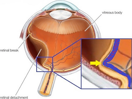
Background: To review cases of branch retinal vein occlusion (BRVO) secondary to rhegmatogenous retinal detachment (RRD) and its surgical management and presume their mechanism.

Methods: Medical records of patients who underwent surgery for RRD between 2015 and 2019 at a single tertiary care center were retrospectively reviewed. New BRVO secondary to RRD or its surgical procedure was diagnosed based on the fundus examination and its clinical course.

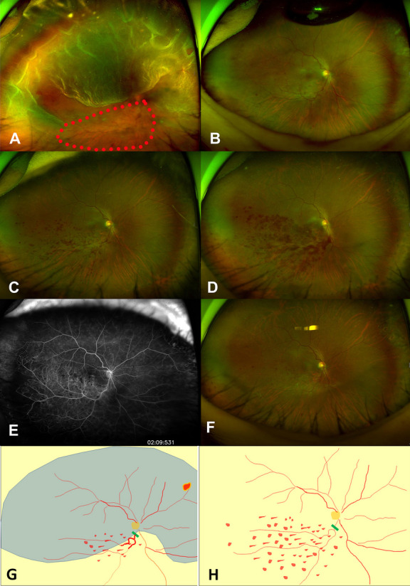
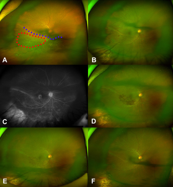
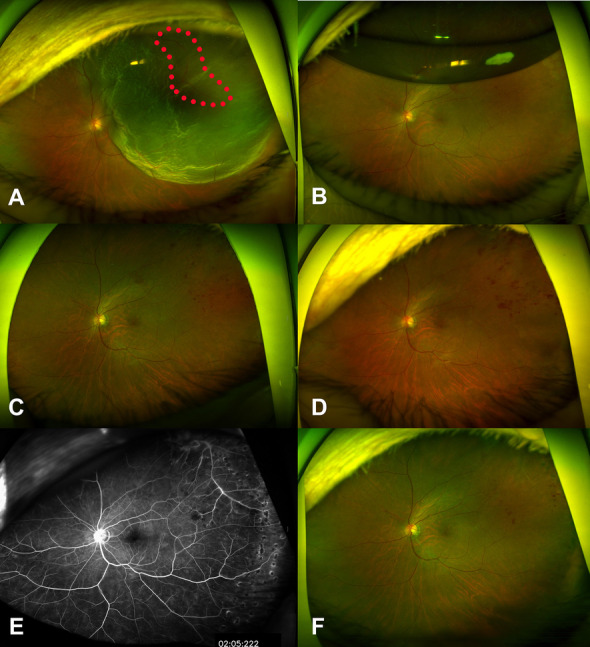
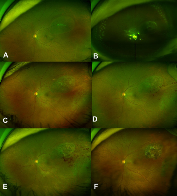
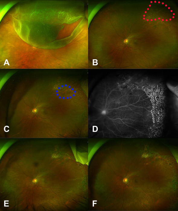
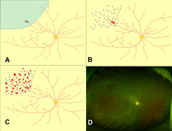
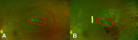
Results: A total of 734 RRD surgeries were performed for five years, and six cases of new BRVOs were noticed in the first year after surgery (incidence was 0.68%: six cases of BRVO / 734 cases of surgical RRD); five cases occurred after vitrectomy, and one occurred after scleral buckling. In three cases, retinal veins were presumed to already be partially occluded related due to a kink of the retinal vein seen before surgery. In the other three cases, the retinal veins were presumed to have incurred damage during vitrectomy.

Conclusion: In the present cohort, RRD or its related procedures caused BRVO within a year of surgery at an incidence of 0.68%. The proposed mechanisms are kinks of the retinal vein on the detached retina and damage to the retinal vein during vitrectomy.

Keywords: Branch retinal vein occlusion; Complication; Rhegmatogenous retinal detachment; Scleral buckling; Vitrectomy.


Conflict of interest statement

The authors declare no competing interests.

Figures

Fig. 1
Fig. 1
Series of fundus photographs of Case 1, a 48-year-old woman who presented with rhegmatogenous retinal detachment of the right eye invading from the 8 o’clock to 4 o’clock positions and involving the whole macula. Preoperatively, a beaded retinal vein with surrounding dot hemorrhage (red dots circle) was present inferior to the macula (A). A fundus photo taken at 10 days postoperatively showed tamponading air and the continued presence of the beaded retinal vein and adjacent dot hemorrhage around it (B). The dot hemorrhage appeared to have worsened in severity by postoperative week 4 (C) and to have worsened further at postoperative week 8 (D). Fluorescein angiography performed at 10 months postoperatively revealed no additional pathology (E). The dot hemorrhage was decreased in the fundus photo taken at 1 year postoperatively (F). A drawing of the preoperative fundus features (Fig. 1A) showed detached retina (blue color) and territory of the occluded retinal vein (G). Green bar indicates the presumed occluded lesion of retinal vein (G). A drawing of fundus features at 4 weeks after the surgery (Fig. 1D) (H)
Fig. 2
Fig. 2
Series of fundus photos of Case 2 a 63-year-old woman who presented with retinal detachment of the right eye invading from the 2 o’clock to 8 o’clock positions of the retina and involving the fovea; a horseshoe tear was also noted at the 7 o’clock position preoperatively and the demarcation line (blue dots) crossed the macula obliquely from the 2 o’clock to 10 o’clock positions (A). The inferior retinal vein appeared tortuous and there was some evidence of dot hemorrhage (red dots circle) in the territory of the tortuous vein (A). Fundus photo taken at 8 weeks postoperatively showed that the detached retina had reattached after scleral buckling (B). The beaded retinal vein looked healthier than before the surgery, but the dot hemorrhage increased (B). Fluorescein angiography taken at 8 weeks postoperatively showed no pathology other than branch retinal vein occlusion and rhegmatogenous retinal detachment (C). The hemorrhage appears to have worsened at 15 weeks postoperatively (D). The hemorrhage appears to have improved in the fundus photo taken at 6 months postoperatively (E) and that taken at 1 year postoperatively (F)
Fig. 3
Fig. 3
Series of fundus photos of Case 3, a 74-year-old woman who presented with rhegmatogenous retinal detachment of the left eye. Preoperative fundus photo showed bullous retinal detachment from the 11 o’clock to 4 o’clock positions involving the fovea and tortuous and beaded veins (red dots circle) were visible in the superotemporal quadrant (A). Fundus photo at 1 week postoperatively showed a retinal hemorrhage in the superotemporal quadrant, partially obscured by tamponading air (B). The hemorrhage increased in the fundus photo at 4 weeks postoperatively (C) and that taken at 9 weeks postoperatively (D). Fluorescein angiography at 9 weeks postoperatively revealed no additional pathology (E). The hemorrhage appeared to have improved in the fundus photo taken at 1 year postoperatively (F)
Fig. 4
Fig. 4
Series of fundus photos of Case 4, a 63-year-old woman who presented with rhegmatogenous retinal detachment of the left eye. A preoperative fundus photo showed localized retinal detachment at the mid-peripheral retina of the superotemporal quadrant and a tear was positioned along the large superior temporal retinal vein and appeared strongly attached to the vitreous (A). The retinectomy site, surrounding fresh laser burns, and tamponading air were visible in the fundus photo on postoperative day one (B). A retinal dot hemorrhage was visible in the temporal periphery at four weeks postoperatively (C) and had progressively worsened at eight (D) and ten (E) weeks postoperatively. The retinal dot hemorrhage was almost completely resolved at one year follow up (F)
Fig. 5
Fig. 5
Series of fundus photos of Case 5, a 64-year-old woman who presented with rhegmatogenous retinal detachment of the left eye. A preoperative fundus photo showed retinal detachment invading the retina from the 10 o’clock to 2 o’clock positions and a retinal tear along the superior temporal large retinal vein and the retinal tear appeared strongly attached to the vitreous and retinal vein (A). Red dots circle indicated the retinectomy lesion. No distinct retinal hemorrhage or abnormality of the retinal circulation was noted at 2 weeks postoperatively (B). Multiple retinal hemorrhages (Blue dots circle) were first noted around the retinectomy site 4 weeks postoperatively (C). Some diffuse leakage, including leakage of the optic disc, was noted on fluorescein angiography at 12 weeks postoperatively (D). The retinal hemorrhage appeared worsened in the fundus photo taken at 18 weeks postoperatively (E). The hemorrhage had almost resolved at 1 year postoperatively (F)
Fig. 6
Fig. 6
Retinal findings of Case 6, a 48-year-old man who presented with rhegmatogenous retinal detachment of the right eye. A drawing of the preoperative fundus features illustrates the observed retinal detachment invading retina from the 10 o’clock to 1 o’clock positions and the retinal tear along the retinal vein in the superotemporal region (A). No retinal hemorrhages were visible at 1 week postoperatively (B). A retinal hemorrhage was present in the superotemporal region at 4 weeks postoperatively (C). The retinal hemorrhage had almost resolved at 18 months postoperatively (D)
Fig. 7
Fig. 7
Proposed mechanism I of retinal vein occlusion after rhegmatogenous retinal detachment (RRD) (cases 1, 2, and 3). Kinking of the retina is common in RRD and causes folding of the retinal vein. Folding of the retinal vein may increase the likelihood of retinal vein stasis, especially in patients with older age, hypertension, or dyslipidemia
Fig. 8
Fig. 8
Proposed mechanism II of retinal vein occlusion after rhegmatogenous retinal detachment (RRD) (case 4, 5, and 6). Fundal photo (Case 4) showed that the vitreous was strongly adherent to a large retinal vein where retinal tears (red dots circle) occurred (A) New branch retinal vein occlusion occurred near this site after surgery. The presumed obstructive lesion (yellow line) was just proximal to the retinectomy site (red dots circle) (B)

Similar articles

References

    1. Kolar P. Risk factors for central and branch retinal vein occlusion: a meta-analysis of published clinical data. J Ophthalmol. 2014;2014:1–5. doi: 10.1155/2014/724780. - DOI - PMC - PubMed
    1. Li J, Paulus YM, Shuai Y, Fang W, Liu Q, Yuan S. New developments in the classification, pathogenesis, risk factors, natural history, and treatment of branch retinal vein occlusion. J Ophthalmol. 2017;2017:1–18. - PMC - PubMed
    1. Jaulim A, Ahmed B, Khanam T, Chatziralli IP. Branch retinal vein occlusion. Retina. 2013;33(5):901–910. doi: 10.1097/IAE.0b013e3182870c15. - DOI - PubMed
    1. Newman-Casey PA, Stem M, Talwar N, Musch DC, Besirli CG, Stein JD. Risk factors associated with developing branch retinal vein occlusion among enrollees in a United States managed care plan. Ophthalmology. 2014;121(10):1939–1948. doi: 10.1016/j.ophtha.2014.04.045. - DOI - PMC - PubMed
    1. Dodson PM, Kubicki AJ, Taylor KG, Kritzinger EE. Medical conditions underlying recurrence of retinal vein occlusion. Br J Ophthalmol. 1985;69(7):493–496. doi: 10.1136/bjo.69.7.493. - DOI - PMC - PubMed