

The role of point-of-care ultrasound in the assessment of pelvic urine leakage and diagnosis of urinoma
- PMID: 38110890
- PMCID: PMC10726508
- DOI: 10.1186/s12245-023-00571-4
The role of point-of-care ultrasound in the assessment of pelvic urine leakage and diagnosis of urinoma
Abstract
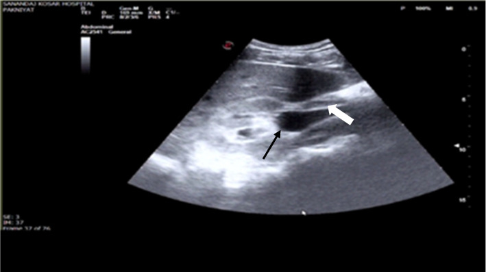
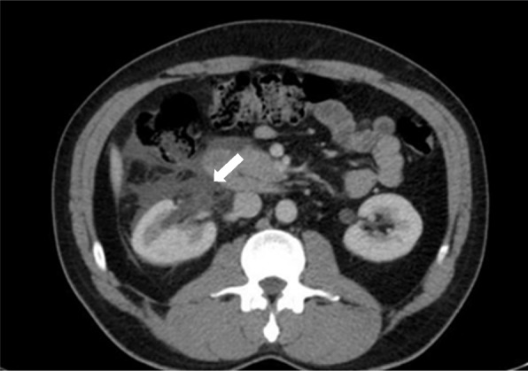
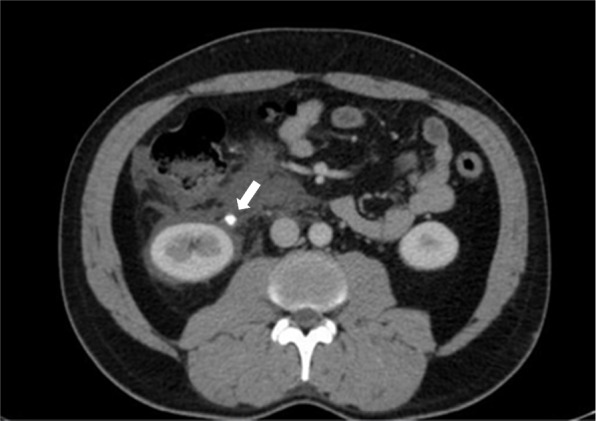
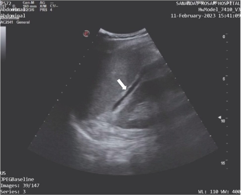
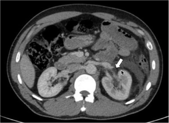
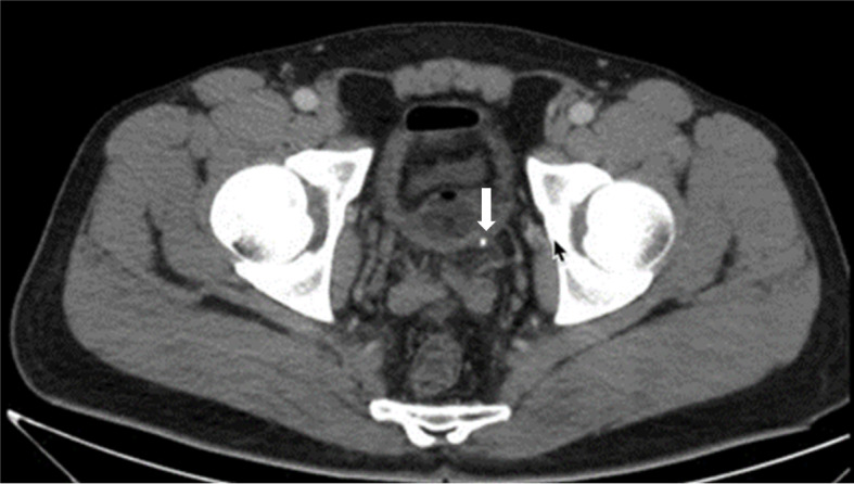
Background: Urinoma, a rare condition resulting from urine leakage due to trauma to the kidney, bladder, or urethra, is typically diagnosed using enhanced computed tomography urogram with delayed imaging. This report presents two cases of urinoma likely caused by overdistention of the renal pelvis following excessive fluid intake and the presence of a ureteral stone.
Case presentation: We present two cases of 36-year-old and 38-year-old patients who presented with flank pain. point-of-care ultrasound (POCUS) played a pivotal role in identifying perinephric fluid in Morrison's space and the splenorenal space, respectively. These ultrasound findings guided further investigations, leading to definitive diagnoses via abdominal pelvic CT scans. Treatment involved prophylactic antibiotics and the successful placement of a double J stent into the renal pelvis over the wire under fluoroscopic guidance, which resulted in significant clinical improvement for both patients.
Conclusions: This study demonstrates the rare occurrence of urinoma from urolithiasis, the use of POCUS in expediting diagnosis and treatment, and the importance of interpreting sonographic images in the correct clinical setting.
Keywords: Case report; Point-of-care ultrasound; Renal pelvis rupture; Urinoma.
© 2023. The Author(s).
Conflict of interest statement
The authors declare no competing interests.
Figures
References
-
- Petrisor O, et al. Perirenal urinomas a complication of the upper urinary tract lithiasis: CT aspects. 2012. European Congress of Radiology-ECR 2012.
LinkOut - more resources
Full Text Sources
