. 2023 Dec 18;21(1):292.
doi: 10.1186/s12915-023-01790-0.

GPRC5C regulates the composition of cilia in the olfactory system

Affiliations

GPRC5C regulates the composition of cilia in the olfactory system

Sneha Bhat et al. BMC Biol. .

Abstract

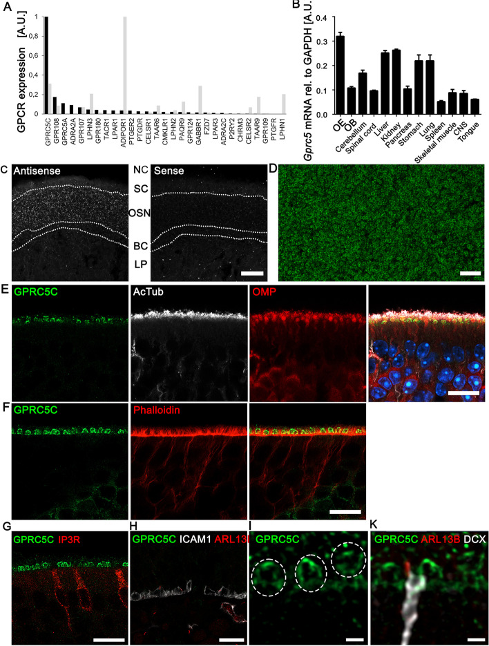
Background: Olfactory sensory neurons detect odourants via multiple long cilia that protrude from their dendritic endings. The G protein-coupled receptor GPRC5C was identified as part of the olfactory ciliary membrane proteome, but its function and localization is unknown.

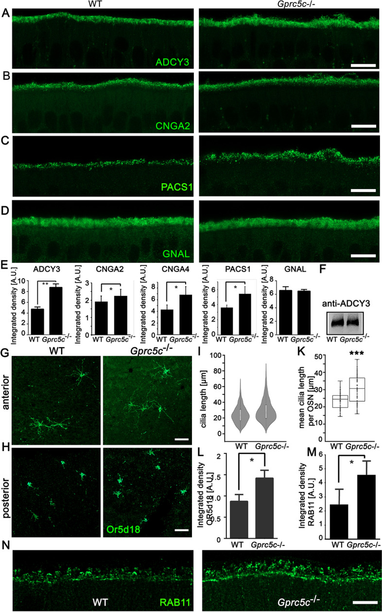
Results: High-resolution confocal and electron microscopy revealed that GPRC5C is located at the base of sensory cilia in olfactory neurons, but not in primary cilia of immature neurons or stem cells. Additionally, GPRC5C localization in sensory cilia parallels cilia formation and follows the formation of the basal body. In closer examination, GPRC5C was found in the ciliary transition zone. GPRC5C deficiency altered the structure of sensory cilia and increased ciliary layer thickness. However, primary cilia were unaffected. Olfactory sensory neurons from Gprc5c-deficient mice exhibited altered localization of olfactory signalling cascade proteins, and of ciliary phosphatidylinositol-4,5-bisphosphat. Sensory neurons also exhibited increased neuronal activity as well as altered mitochondrial morphology, and knockout mice had an improved ability to detect food pellets based on smell.

Conclusions: Our study shows that GPRC5C regulates olfactory cilia composition and length, thereby controlling odour perception.

Keywords: Cilia; GPRC5C; Olfactory; Sensory; Smell.


Conflict of interest statement

The authors declare that they have no competing interests.

Figures

Fig. 1
Fig. 1
GPRC5C is expressed in dendritic knobs of OSNs. A Relative expression levels of GPCRs in cilia preparations of OSNs (black bars) and in next-generation sequencing results of FACS sorted OSNs (grey bars), shown are relative intensity or FPKM levels, each normalized to the most abundant GPCR in both data sets. Shown are non-olfactory GPCRs that have been identified with proteomic methods. B qPCR analysis of Gprc5c mRNA expression across different tissues in adult mice. CNS is brain excluding olfactory bulb (OB) (n = 3, individual data values Additional file 2). C Confocal images of adult OE cryosection upon FISH of DIG-labelled probes for GPRC5C (antisense and sense). Strong GPRC5C mRNA expression is detected in the neuronal cell layer (OSN). The apical sustentacular cell layer (SC) and basal cell layers (BC) do not express GPRC5C mRNA. Dotted lines demarcate the different cell layers. NC: Nasal cavity, LP: Lamina propria. Sense control showed no labelling. Scale bar: 20 µm. D En face preparation of the OE showing expression of GPRC5C in all dendritic knobs. Confocal images of adult OE coronal cryosection immunolabelled for E GPRC5C (green), acetylated-tubulin (AcTub) (white), OMP (red), and overlay along with DAPI (blue) showing localization in dendritic knobs of olfactory neuron F GPRC5C (green) and Phalloidin (red) showing GPRC5C + structures embedded in between sustentacular cell microvilli. G GPRC5C (green) and IP3R (red) showing that GPRC5C is not localized to the apical surface of microvillar cells. H Absence of GPRC5C (labelled in green) in ICAM-positive (white) HBCs, primary cilia are labelled with ARL13B (red), white line marks the basal lamina. I GPRC5C (green)-positive dendritic knobs, single-channel image for overlay shown in K. K ARL13B (red)-positive primary cilia are present on immature OSNs labelled with DCX (white), but not on GPRC5C (green) labelled dendritic knobs. Scale bars E–H 10 µm and I–K 1 µm
Fig. 2
Fig. 2
Localization of GPRC5C in the OE during regeneration of the OE. A Confocal images of OE cryosections of regenerating OE (3dpi, 14dpi and 28dpi), immunolabelled for GPRC5C (green) and GAP43 (red, left panel) and OMP (red, right panel). At 3dpi, no neurons were present and GPRC5C staining in the OE was not apparent. Dendritic knobs with GPRC5C expression can be seen at 14dpi and as regeneration of the neurons progresses, more knobs with GPRC5C staining are observed at 28dpi. B Quantification of GPRC5C positive knobs at 3-, 14- and 28-days post MMZ injection, counted in projections of confocal stacks of 16-μm thickness (n = 3 animals per group, individual data values Additional file 2, Student’s t test, error bars represent SEM, ***p < 0.001). C Confocal images of OE at 14dpi and 28dpi immunolabelled for GPRC5C (green) and γ-tubulin (red, left panel) and acetylated-tubulin (red, right panel). At 14dpi, GPRC5C + dendritic knobs have few newly formed acetylated-tubulin stained cilia, and at 28dpi, the amount of cilia increased. D Quantification of GPRC5C and γ-tubulin co-localization. Ca. 50% of the γ-tubulin + knobs express GPRC5C at 14dpi, at 28dpi ca. 80% of the dendritic knobs co-express GPRC5C and gamma-tubulin. Knobs were counted in projections of confocal stacks of 16-μm thickness, percentage co-expression was analysed using Student’s t test (n = 3 animals per group, individual data values Additional file 2, error bars represent SEM, **p < 0.01). E Z-stack projections of confocal images of WT and Gprc5c−/− adult mouse OE cryosections showing comparable ARL13B staining. F Quantifications of staining intensity of ARL13B. Student’s t test showed no significant difference (n = 3, individual data values Additional file 2). Scale bars 10 µm
Fig. 3
Fig. 3
Gprc5c/ mice show altered ciliary morphology. A Bright-field image of ß-Gal staining of cryosections of Gprc5c−/− adult mouse OE. ß-Gal staining is confined to the neuronal layer in the adult OE. Dotted line represents basal lamina. B Lower magnification overview image. C Confocal images of adult OE cryosection upon FISH of DIG-labelled probes for Gprc5c (antisense). Gprc5c mRNA was detected in WT, but not in Gprc5c−/− OE. Dotted line represents basal lamina. Transmission electron micrographs of ultrathin sections OE of WT (D) and Gprc5c−/− (E). Gprc5c−/− OE depicts an increased thickness of the ciliary layer. F Scanning electron micrographs of WT OE appears organized with dendritic knobs evenly embedded in the surrounding ciliary network. G Higher magnification. H Gprc5c−/− OE appears disorganized and has atypical cilia. I Higher magnification. K Cilium with bending of the distal end. L Clumped cilia. M Extracellular vesicles attached to cilia. N Quantification of irregular cilia. Mann–Whitney test showed significant increase in number of irregular cilia in Gprc5c−/− OE (n = 4, individual data values Additional file 2, error bars represent SEM, *p < 0.05). O Staining of acetylated-tubulin (acTub) as marker for the axoneme of the cilia shows an increase in staining in Gprc5c−/− OE. P Quantification of staining intensity followed by Student’s t test revealed a significant increase of acetylated-tubulin in Gprc5c−/− OE (n = 4, individual data values Additional file 2, error bars represent SEM, *p < 0.05). Q Western blot analysis of whole OE preparations, acetylated-tubulin band at 52 kDa, levels were comparable between WT and Gprc5c−/− OE, equal loading was controlled by actin. Scale bars A, C, O 10 µm; B 500 µm: D, E, F, H 2 μm; G, I 400 nm; K, L, M 200 nm
Fig. 4
Fig. 4
GPRC5C is localized at the ciliary gate. Confocal images of coronal sections (A) and en face preparation (B) adult mouse OE, immunolabelled for GPRC5C (green) and γ-tubulin (red) show punctate localization of GPRC5C along the membrane of the dendritic knob in a ring-like manner surrounding a γ-tubulin cluster that is localized in the lumen of the dendritic knob. Arrow indicates an individual dendritic knob. C Airyscan image with deconvolution processing of immunolabelling for GPRC5C (green), γ-tubulin (red) and acetylated-tubulin (white). GPRC5C is localized in the region between the basal body and the cilia. Arrow indicates an individual dendritic knob. D Immunogold labelling of freeze-fracture replica of the OE with GPRC5C antibody. Clusters of immunogold particles localized to the base of the cilia (black arrows). Higher magnification images show a distinct localization of GPRC5C at the ciliary necklace region, characterized by rows of ciliary proteins (arrow). E Quantitative analysis of anti-GPRC5C immunogold labellings of freeze-fractured plasma membranes of proximal and distal cilia. Proximal dendrite, n = 50 ROIs; distal dendrite, n = 40 ROIs. F, G Confocal images of FBF1 (green) and γ-tubulin (red), H, I MKS3 (green) and γ-tubulin (red). The localization pattern of FBF1 and MKS3 in the dendritic knob is similar to that of GPRC5C, all showing punctate staining along the membrane of the knob surrounding the γ-tubulin staining in the core. K Quantification of MKS3 staining intensity. Significance obtained upon performing Student’s t test (n = 3, individual data values Additional file 2, error bars represent SEM, *p < 0.05). L, M Confocal images of WT and Gprc5c−/− OE immunolabelled MKS3 showing increased staining intensity and altered pattern of staining in Gprc5c− −dendritic knobs. Scale bars A, B, G, I, M 2 µm; C 1 µm; D 200 nm (right), 100 nm (left), F, H 10 µm; L 5 µm
Fig. 5
Fig. 5
Localization of ciliary markers in WT and Gprc5c−/− OE. Z-stack projections of confocal images of WT and Gprc5c−/− adult mouse OE cryosections immunolabelled for A ADCY3, B CNGA2, C PACS1 and D GNAL. Scale bar 10 µm. While GNAL (Gαolf) staining is comparable between WT and Gprc5c− −ADCY3, CNGA2 and PACS1 ≥ is increased in Gprc5c−/− cilia. E Quantifications of staining intensity of ciliary markers. Student’s t test showed significant increase in staining intensity for ADCY3, CNGA2, CNGA4, PACS1 and GNAL (n = 3–4, individual data values Additional file 2, error bars represent SEM, *p < 0.05, **p < 0.05). F Western blot analysis of ADCY3 expression in whole OE preparations, band slightly above 180 kDa, equal loading was controlled by actin. G Confocal images of en face preparation of septal OE of adult WT and Gprc5c−/− mice immune-labelled for Or5d18, depicting the anterior region. Scale bar: 10 μm. H In the posterior region, cilia are in general shorter compared to the anterior region. I Violin plots of length of Or5d18 cilia in WT and Gprc5c−/− mice. K Quantification of the length of Or5d18 + cilia per OSN. Student’s t test showed significant increase in Gprc5c−/− OE (n ≥ 50 knobs, error bars represent SEM, ***p < 0.001). L Quantification of staining intensity of posterior mOR-EG neurons in WT and Gprc5c−/− OE. Student’s t test showed significant increase in Gprc5c−/− OE (n = 3, individual data values Additional file 2, error bars represent SEM, * p < 0.05). M Increased localization of RAB11-positive structures in the dendritic knobs of Gprc5c−/− mice (n = 4 animals per group, individual data values Additional file 2, Student’s t test, error bars represent SEM, *p < 0.05). N Z-stack projections of confocal images of WT and Gprc5c−/− OE cryosections immunolabelled for RAB11. Scale bar 10 µm
Fig. 6
Fig. 6
Redistribution of ciliary phospholipids in Gprc5c−/− OE. Confocal images of WT and Gprc5c−/− adult mouse OE cryosections immunolabelled for A PtdIns(4,5)P2 and B PtdIns4P. PtdIns(4,5)P2 staining is restricted to the dendritic knobs in WT OE whereas, Gprc5c−/− OE shows several prominent puncta in the ciliary layer. PtdIns4P on the other hand showed a notable reduction in the Gprc5c−/− cilia. C Quantification of PtdIns(4,5)P2 staining intensity. Student’s t test showed significant increase in staining intensity (n = 3, individual data values Additional file 2, error bars represent SEM, **p < 0.01). D Average expression and percent of respective OE cell types expressing different (putative) 5-phosphatases, OSNs are highlighted, data taken from [55]. HBC-horizontal basal cell, GBC-globose basal cell, INP-immediate neuronal progenitor cell, iOSN-immature olfactory neuron, OSN-mature olfactory neuron, MV-microvillar cell, SUS-sustentacular cell. Scale bars: A, B 10 µm
Fig. 7
Fig. 7
Effects of GPRC5C knockout on neuronal activity and behaviour. A Quantification for buried food test. Gprc5c−/− mice exhibit a significantly reduced latency (WT 395.4 ± 63.66 s, Gprc5c−/− 226.5 ± 36.49 s) to find buried food compared to WT mice, indicating an altered behavioural response (n = 18 animals per group, Student’s t test, error bars represent SEM, * p < 0.05). B Tile scans of the septum of the OE stained for Na/K + ATPase, both images were taken with the same microscope settings. C Z-stack projections of confocal images of WT and Gprc5c−/− adult mouse OE cryosections immunolabelled for PS6. D Quantification of staining intensity of PS6 neurons in WT and Gprc5c−/− OE Student’s t test showed significant increase in Gprc5c−/− OE (n = 3, individual data values Additional file 2, error bars represent SEM, *p < 0.05). E Quantification of number of PS6 neurons. Student’s t test showed significant increase in Gprc5c− −E (n = 3, individual data values Additional file 2, error bars represent SEM, ***p < 0.001). F Quantification of the number of mitochondria per dendritic knob (n = 155 WT, n = 162 Gprc5c−/−). Mann–Whitney test shows no significant difference. G TEM of ultrathin sections of WT and Gprc5c−/− OE depicting altered mitochondria in dendritic knobs of Gprc5c−/− OE. Scale bar 400 nm. H Quantification of MT-CO1 staining as mitochondrial marker. Student’s t test shows significant reduction in staining intensity of MT-CO1 in dendritic knob layer of Gprc5c−/− OE (n = 3, individual data values Additional file 2, error bars represent SEM, **p < 0.01). I Z-stack projections of confocal images of WT and Gprc5c−/− adult mouse OE cryosections immunolabelled for MTCO1 (red) showing reduced staining in dendritic knobs of Gprc5c−/− OE. Scale bar 10 μm

References

    1. Firestein S. How the olfactory system makes sense of scents. Nature. 2001;413(6852):211–218. doi: 10.1038/35093026. - DOI - PubMed
    1. McClintock TS, Khan N, Xie C, Martens JR. Maturation of the olfactory sensory neuron and its cilia. Chem Senses. 2020;45(9):805–822. doi: 10.1093/chemse/bjaa070. - DOI - PMC - PubMed
    1. Jenkins PM, McEwen DP, Martens JR. Olfactory cilia: linking sensory cilia function and human disease. Chem Senses. 2009;34(5):451–464. doi: 10.1093/chemse/bjp020. - DOI - PMC - PubMed
    1. Menco BP. Ciliated and microvillous structures of rat olfactory and nasal respiratory epithelia. A study using ultra-rapid cryo-fixation followed by freeze-substitution or freeze-etching. Cell Tissue Res. 1984;235(2):225–41. doi: 10.1007/BF00217846. - DOI - PubMed
    1. Menco BP. Ultrastructural aspects of olfactory signaling. Chem Senses. 1997;22(3):295–311. doi: 10.1093/chemse/22.3.295. - DOI - PubMed

Publication types

Substances

LinkOut - more resources