. 2023 Dec 18;30(1):96.
doi: 10.1186/s12929-023-00987-3.

A novel mucosal bivalent vaccine of EV-A71/EV-D68 adjuvanted with polysaccharides from Ganoderma lucidum protects mice against EV-A71 and EV-D68 lethal challenge

Affiliations

A novel mucosal bivalent vaccine of EV-A71/EV-D68 adjuvanted with polysaccharides from Ganoderma lucidum protects mice against EV-A71 and EV-D68 lethal challenge

Yu-Li Lin et al. J Biomed Sci. .

Abstract

Background: Human enteroviruses A71 (EV-A71) and D68 (EV-D68) are the suspected causative agents of hand-foot-and-mouth disease, aseptic meningitis, encephalitis, acute flaccid myelitis, and acute flaccid paralysis in children. Until now, no cure nor mucosal vaccine existed for EV-A71 and EV-D68. Novel mucosal bivalent vaccines are highly important for preventing EV-A71 and EV-D68 infections.

Methods: In this study, formalin-inactivated EV-A71 and EV-D68 were used as antigens, while PS-G, a polysaccharide from Ganoderma lucidum, was used as an adjuvant. Natural polysaccharides have the characteristics of intrinsic immunomodulation, biocompatibility, low toxicity, and safety. Mice were immunized intranasally with PBS, EV-A71, EV-D68, or EV-A71 + EV-D68, with or without PS-G as an adjuvant.

Results: The EV-A71 + EV-D68 bivalent vaccine generated considerable EV-A71- and EV-D68-specific IgG and IgA titres in the sera, nasal washes, saliva, bronchoalveolar lavage fluid, and feces. These antibodies neutralized EV-D68 and EV-A71 infectivity. They also cross-neutralized infections by different EV-D68 and EV-A71 sub-genotypes. Furthermore, compared with the PBS group, EV-A71 + EV-D68 + PS-G-vaccinated mice exhibited an increased number of EV-D68- and EV-A71-specific IgA- and IgG-producing cells. In addition, T-cell proliferative responses, and IFN-γ and IL-17 secretion in the spleen were substantially induced when PS-G was used as an adjuvant with EV-A71 + EV-D68. Finally, in vivo challenge experiments demonstrated that the immune sera induced by EV-A71 + EV-D68 + PS-G conferred protection in neonate mice against lethal EV-A71 and EV-D68 challenges as indicated by the increased survival rate and decreased clinical score and viral RNA tissue expression. Taken together, all EV-A71/EV-D68 + PS-G-immunized mice developed potent specific humoral, mucosal, and cellular immune responses to EV-D68 and EV-A71 and were protected against them.

Conclusions: These findings demonstrated that PS-G can be used as a potential adjuvant for EV-A71 and EV-D68 bivalent mucosal vaccines. Our results provide useful information for the further preclinical and clinical development of a mucosal bivalent enterovirus vaccine against both EV-A71 and EV-D68 infections.

Keywords: Acute flaccid myelitis; Acute flaccid paralysis; Adjuvant; Enterovirus A71; Enterovirus D68; Ganoderma lucidum polysaccharide; Intranasal; Mucosal vaccine.


Conflict of interest statement

The authors declare that they have no competing interest.

Figures

Fig. 1
Fig. 1
Long-term memory immunity induced by inactivated EV-A71 or EV-D68 mucosal vaccines in mice. Mice were intranasally immunized with PBS, formalin-inactivated EV-A71 (2.5 μg/mouse), or EV-D68 (2.5 μg/mouse) thrice at 3-week intervals. a Immunization schedules for the EV-A71 mucosal vaccine. Serum and faecal samples were collected from immunized mice at weeks 8, 11, 14, 22, and 24. The levels of EV-A71-specific IgG and IgA in the serum (b, c) and feces (d, e) were measured using ELISA. f Neutralization titre against EV-A71 C2 infection in the serum of immunized mice. g Immunization schedules for the EV-D68 mucosal vaccine. Serum, feces, and saliva samples were collected from immunized mice at weeks 8, 11, 14, 25, and 27. The levels of EV-D68-specific IgG and IgA in the serum (h, i), feces (j, k), and saliva (l, m) were measured using ELISA. n Neutralization titre against EV-D68 1788 infection in the serum of immunized mice. *p < 0.05; **p < 0.01; ***p < 0.001
Fig. 2
Fig. 2
Effect of the EV-A71 + EV-D68 bivalent vaccine on EV-D68- and EV-A71-specific IgG and IgA and neutralization titres against infection by different enteroviruses in immunized mice. Mice were intranasally immunized with PBS, formalin-inactivated EV-A71 (2.5 μg/mouse), formalin-inactivated EV-D68 (2.5 μg/mouse), and combined formalin-inactivated EV-A71 (2.5 μg/mouse) and EV-D68 (2.5 μg/mouse) thrice at 3-week intervals. a Three-dose immunization schedules. b The levels of EV-D68-specific IgA and IgG and c EV-A71-specific IgA and IgG in the saliva, nasal wash, BALF, and feces of mice after the third immunization were measured using ELISA. d The levels of EV-D68-specific IgG and IgA and e EV-A71-specific IgG and IgA in the sera of mice after the third intranasal immunization were measured using ELISA. Sera were serially diluted (23–212), mixed with f EV-D68 and g EV-A71 virus, and used to infect RD cells. After 4 d, the the highest dilution that resulted in the virus producing no cytopathic effect was considered to be the neutralization titre. All data are expressed as the mean ± SEM. *p < 0.05, **p < 0.01, ***p < 0.001
Fig. 3
Fig. 3
Effect of the EV-A71 + EV-D68 bivalent vaccine on the number of antibody-secreting cells (ASCs), T-cell proliferation, and cytokine release in splenocytes. Mice were intranasally immunized with PBS, formalin-inactivated EV-A71 (2.5 μg/mouse) alone, formalin-inactivated EV-D68 (2.5 μg/mouse) alone, and combined formalin-inactivated EV-A71 (2.5 μg/mouse) with EV-D68 (2.5 μg/mouse) thrice at 3-week intervals. Spleen was isolated 2 weeks after the third immunization, and then the numbers of a EV-D68-specific IgG and IgA ASCs and b EV-A71-specific IgG and IgA ASCs were measured using ELISPOT. c, d Splenocytes from immunized mice were harvested and cultured in medium containing 10 μg/mL heat-inactivated EV-D68 or EV-A71. After culture for 5 d, proliferation was measured as [3H]-thymidine incorporation in splenocytes. Culture supernatants of splenocytes were analysed for levels of IFN-γ, IL-17, and IL-4 after 2 d in response to heat-inactivated EV-D68 or EV-A71. Thymidine uptake was determined by harvesting cells and using a scintillation counter to measure the level of incorporation (as counts per minute; cpm). All data are expressed as the mean ± SEM. *p < 0.05, **p < 0.01, ***p < 0.001
Fig. 4
Fig. 4
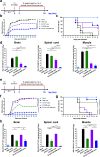
Protective efficacy of the inactivated EV-A71 + EV-D68 vaccine in the EV-D68 or EV-A71 infection mouse model. The passive sera transfer and virus challenge schedules. Briefly, 2-d-old ICR mice were i.p. injected with 20 μL anti-PBS, anti-EV-A71, anti-EV-D68, or anti-EV-A71 + EV-D68 sera. After 6 h, the suckling mice were intracerebrally (i.c.) administered with RD medium, a EV-D68 (CDC_NO 2016-05298), or e EV-A71 (MP4). Mock mice were administered medium only. Mice were monitored daily for b, f clinical score and c, g survival rate for 14 d following infection. Clinical scores were graded as described in “Methods”. d, h Brain, spinal cord, and muscle were collected after infection, and viral loads within the indicated tissues were determined using real-time quantitative PCR (qRT-PCR). The levels of expression were normalized to those of GAPDH. Data are presented as the mean ± SEM. *p < 0.05, **p < 0.01, ***p < 0.001
Fig. 5
Fig. 5
Effect of the EV-A71 + EV-D68 bivalent vaccine with or without PS-G or CpG as adjuvant on the levels of EV-D68-specific and EV-A71-specific IgA and IgG in the saliva, nasal wash, BALF, and feces of mice. Mice were intranasally immunized with PBS, formalin-inactivated EV-A71 (2.5 μg/mouse) and EV-D68 (2.5 μg/mouse), and formalin-inactivated EV-A71 (2.5 μg/mouse) and EV-D68 (2.5 μg/mouse) combined with PS-G (10 μg/mouse) or CpG (20 μg/mouse) as adjuvant thrice at 3-week intervals. The levels of a EV-D68-specific IgA and IgG, and b EV-A71-specific IgA and IgG in the saliva, nasal wash, BALF, and feces of mice after the third immunization were measured using ELISA. All data are expressed as the mean ± SEM. *p < 0.05, **p < 0.01, ***p < 0.001
Fig. 6
Fig. 6
Effect of the EV-A71 + EV-D68 bivalent vaccine with or without PS-G or CpG as adjuvant on the levels of EV-D68- and EV-A71-specific IgG and IgA, and neutralization titre against infection by different enteroviruses in the sera of mice. The levels of a EV-D68-specific IgG and IgA and b EV-A71-specific IgG and IgA in the sera of mice after the third intranasal immunization were measured using ELISA. Sera were serially diluted (23–212), mixed with c EV-D68 and d EV-A71 virus, and used to infect RD cells. After 4 d, the highest dilution that resulted in the virus producing no cytopathic effect was considered to be the neutralization titre. All data are expressed as the mean ± SEM. *p < 0.05, **p < 0.01, ***p < 0.001
Fig. 7
Fig. 7
Effect of the EV-A71 + EV-D68 bivalent vaccine with or without PS-G as adjuvant on the number of antibody-secreting cells (ASCs), T-cell proliferation, and cytokine release in splenocytes. Mice were intranasally immunized with PBS, formalin-inactivated EV-A71 (2.5 μg/mouse) and EV-D68 (2.5 μg/mouse), and formalin-inactivated EV-A71 (2.5 μg/mouse) and EV-D68 (2.5 μg/mouse) combined with PS-G (10 μg/mouse) as adjuvant thrice at 3-week intervals. Spleen was isolated at 2 weeks after the third immunization, and then the numbers of a EV-D68-specific IgG and IgA ASCs and b EV-A71-specific IgG and IgA ASCs were measured using ELISPOT. c, d Splenocytes from immunized mice were harvested and cultured in medium containing 10 μg/mL heat-inactivated EV-D68 or EV-A71. After culture for 5 d, proliferation was measured as [3H]-thymidine incorporation in splenocytes. Culture supernatants of splenocytes were analysed for the levels of IFN-γ, IL-17, and IL-4 after 2 d in response to heat-inactivated EV-D68 or EV-A71. e, f Information on IFN-γ and IL-17 production by CD4+ and CD8+ T-cells in the spleen were obtained from all mice. Splenocytes were collected from mice immunized with PBS, formalin-inactivated EV-A71 + EV-D68, or PS-G-adjuvanted EV-A71 + EV-D68 and cultured in RPMI medium containing 10 μg/mL of heat-inactivated EV-A71 or EV-D68. Cells were cultured for 5 d, and the levels of IFN-γ and IL-17 produced by CD4+ and CD8+ T-cells were measured by flow cytometry. All data are expressed as the mean ± SEM. *p < 0.05, **p < 0.01, ***p < 0.001
Fig. 8
Fig. 8
Protective efficacy of the inactivated EV-A71 + EV-D68 vaccine with or without PS-G as adjuvant in the EV-D68 or EV-A71 infection mouse model. The passive sera transfer and virus challenge schedules are shown. Briefly, 2-d-old ICR mice were i.p. injected with 20 μL anti-PBS, anti-EV-A71 + EV-D68, or anti-EV-A71 + EV-D68 + PS-G sera. After 6 h, the suckling mice were intracerebrally (i.c.) administered with RD medium, a EV-D68 (CDC_NO 2016-05298), or f EV-A71 (MP4). Mock mice were given medium only. b, g Representative images showing symptoms of healthy mice in the EV-A71 + EV-D68 or EV-A71 + EV-D68 + PS-G immunized group and mice with limb paralysis in the PBS-immunized group on day 9 post-infection with EV-D68 or EV-A71. Mice were monitored daily for c, h clinical score and d, i survival rates for 14 d following infection. Clinical scores were graded as described in “Methods”. e, j Brain, spinal cord, and muscle were collected after infection, and viral loads within the indicated tissues were determined using real-time quantitative PCR (qRT-PCR). The levels of expression were normalized to those of GAPDH. All data are expressed as the mean ± SEM. *p < 0.05, **p < 0.01, ***p < 0.001

Similar articles

Cited by

References

    1. Oberste MS, Maher K, Schnurr D, Flemister MR, Lovchik JC, Peters H, et al. Enterovirus 68 is associated with respiratory illness and shares biological features with both the enteroviruses and the rhinoviruses. J Gen Virol. 2004;85:2577–2584. doi: 10.1099/vir.0.79925-0. - DOI - PubMed
    1. Liu Y, Sheng J, Fokine A, Meng G, Shin W-H, Long F, et al. Structure and inhibition of EV-D68, a virus that causes respiratory illness in children. Science. 2015;347:71–74. doi: 10.1126/science.1261962. - DOI - PMC - PubMed
    1. Yuan J, Shen L, Wu J, Zou X, Gu J, Chen J, et al. Enterovirus A71 proteins: structure and function. Front Microbiol. 2018;9:286. doi: 10.3389/fmicb.2018.00286. - DOI - PMC - PubMed
    1. Oermann CM, Schuster JE, Conners GP, Newland JG, Selvarangan R, Jackson MA. Enterovirus D68. A focused review and clinical highlights from the 2014 U.S. Outbreak. Ann Am Thorac Soc. 2015;12:775–781. doi: 10.1513/AnnalsATS.201412-592FR. - DOI - PubMed
    1. Schmidt NJ, Lennette EH, Ho HH. An apparently new enterovirus isolated from patients with disease of the central nervous system. J Infect Dis. 1974;129:304–309. doi: 10.1093/infdis/129.3.304. - DOI - PubMed

Supplementary concepts