. 2023 Dec 18;23(1):126.
doi: 10.1186/s40644-023-00637-9.

Pancreas CT assessment for pancreatic ductal adenocarcinoma resectability: effect of tube voltage and slice thickness on image quality and diagnostic performance

Affiliations

Pancreas CT assessment for pancreatic ductal adenocarcinoma resectability: effect of tube voltage and slice thickness on image quality and diagnostic performance

Dong Ho Lee et al. Cancer Imaging. .

Abstract

Objectives: To assess the resectability of pancreatic ductal adenocarcinoma (PDAC), the evaluation of tumor vascular contact holds paramount significance. This study aimed to compare the image quality and diagnostic performance of high-resolution (HR) pancreas computed tomography (CT) using an 80 kVp tube voltage and a thin slice (1 mm) for assessing PDAC resectability, in comparison with the standard protocol CT using 120 kVp.

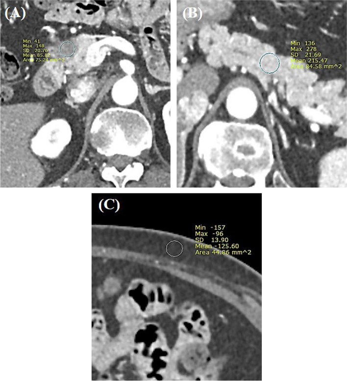
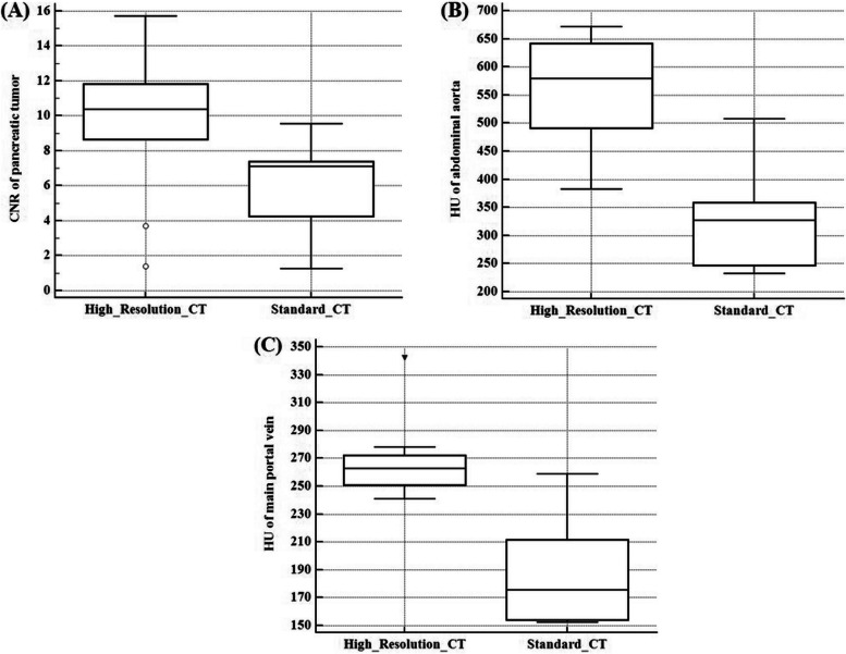
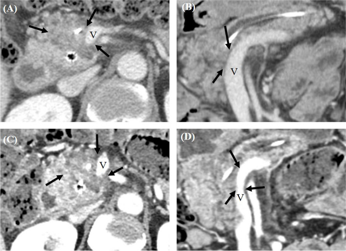
Methods: This research constitutes a secondary analysis originating from a multicenter prospective study. All participants underwent both the standard protocol pancreas CT using 120 kVp with 3 mm slice thickness (ST) and HR-CT utilizing an 80 kVp tube voltage and 1 mm ST. The contrast-to-noise ratio (CNR) between parenchyma and tumor, along with the degree of enhancement of the abdominal aorta and main portal vein (MPV), were measured and subsequently compared. Additionally, the likelihood of margin-negative resection (R0) was evaluated using a five-point scale. The diagnostic performance of both CT protocols in predicting R0 resection was assessed through the area under the receiver operating characteristic curve (AUC).

Results: A total of 69 patients (37 males and 32 females; median age, 66.5 years) were included in the study. The median CNR of PDAC was 10.4 in HR-CT, which was significantly higher than the 7.1 in the standard CT (P=0.006). Furthermore, HR-CT demonstrated notably higher median attenuation values for both the abdominal aorta (579.5 HU vs. 327.2 HU; P=0.002) and the MPV (263.0 HU vs. 175.6 HU; P=0.004) in comparison with standard CT. Following surgery, R0 resection was achieved in 51 patients. The pooled AUC for HR-CT in predicting R0 resection was 0.727, slightly exceeding the 0.699 of standard CT, albeit lacking a significant statistical distinction (P=0.128).

Conclusion: While HR pancreas CT using 80 kVp offered a notably greater degree of contrast enhancement in vessels and a higher CNR for PDAC compared to standard CT, its diagnostic performance in predicting R0 resection remained statistically comparable.

Keywords: High-resolution pancreas CT; Pancreatic ductal adenocarcinoma; Resectability assessment.


Conflict of interest statement

Jeong Min Lee: Grant from Bayer, Bracco, Canon, Central Medical Servis, Dongkuk Pharma, GE Healthcare, Guerbet, Philips Healthcare, Samsung Medison, Siemens Healthineers; Consulting fees from Sansumg medison; Honoraria from Bayer, Claripi, GE Healthcare, Guerbet, Philips Healthcare, Samsung Medison, Siemens Healthineers.

Dong Ho Lee: Research Grants from Canon Medical Systems.

Other authors declared no competing interests.

Figures

Fig. 1
Fig. 1
Assessment of CNR for pancreatic ductal adenocarcinoma in a 56-year-old female patient. A An arterial phase axial contrast-enhanced CT image from the high-resolution protocol reveals a 2 cm hypo-attenuating lesion with a mean Hounsfield Unit (HU) value of 85.8. B The mean HU value for the pancreatic parenchyma was 215.47. C Noise level, characterized by the mean standard deviation of subcutaneous fat attenuation, is measured at three distinct locations (one shown here), yielding a value of 13.5. From these measurements, the CNR for the pancreatic ductal adenocarcinoma is calculated to be 9.6
Fig. 2
Fig. 2
Quantitative analysis results for (A) the CNR of pancreatic ductal adenocarcinoma; (B) the attenuation value of the abdominal aorta; and (C) the attenuation value of the main portal vein. The central box represents the interquartile range while the middle line represents the median value
Fig. 3
Fig. 3
CT images obtained from a 79-year-old man with pancreatic ductal adenocarcinoma in the pancreas head. A Arterial phase axial contrast enhanced CT image (window width, 400HU; window level, 40HU) acquired through the standard protocol showed a 1.5 cm hypo-attenuating lesion in the pancreas head (arrow) abutting the SMV. The calculated CNR of pancreatic tumor was 7.4. B Arterial phase axial contrast enhanced CT image (window width, 400HU; window level, 40HU) acquired through the high-resolution protocol also revealed a 1.5 cm hypo-attenuating lesion (arrow) with calculated CNR of pancreatic tumor of 15.7. The abutment of SMV was also noted. Upon reviewing these imaging findings, all three readers classified this patient as having resectable pancreatic ductal adenocarcinoma on both CT protocols. Nevertheless, surgical exploration unveiled unexpected peritoneal seeding, making the tumor unresectable
Fig. 4
Fig. 4
CT images from a 57-year-old male with pancreatic ductal adenocarcinoma in the pancreas head. A Portal venous phase axial contrast-enhanced CT image (window width, 400 HU; window level, 40 HU) obtained using the standard protocol displaying a 3.5 cm hypo-attenuating lesion in the pancreas head (arrows) abutting both the SMV and SMA. B Portal venous phase coronal CT image obtained through the standard protocol also revealed abutment of the pancreatic ductal adenocarcinoma to the SMV. Based on these imaging findings, all three reviewers initially categorized this patient as having borderline resectable pancreatic ductal adenocarcinoma. C Using the high-resolution protocol, a portal venous phase axial contrast-enhanced CT image (window width, 400 HU; window level, 40 HU) also identifies the 3.5 cm hypo-attenuating lesion (highlighted by an arrow) abutting the SMV and SMA. D Notably, the high-resolution coronal CT image reveals luminal narrowing of the SMV due to tumor encasement (indicated by arrows) with an involved length of 2.7 cm. Based on this, two out of the three reviewers reclassified the patient's condition as locally advanced pancreatic ductal adenocarcinoma. While a surgical resection was undertaken, the post-operative histopathologic examination confirmed an R1 resection

References

    1. De Angelis R, Sant M, Coleman MP, Francisci S, Baili P, Pierannunzio D, et al. Cancer survival in Europe 1999–2007 by country and age: results of EUROCARE–5-a population-based study. Lancet Oncol. 2014;15(1):23–34. doi: 10.1016/S1470-2045(13)70546-1. - DOI - PubMed
    1. Park SJ, Jang S, Han JK, Kim H, Kwon W, Jang JY, et al. Preoperative assessment of the resectability of pancreatic ductal adenocarcinoma on CT according to the NCCN Guidelines focusing on SMA/SMV branch invasion. Eur Radiol. 2021;31(9):6889–97. doi: 10.1007/s00330-021-07847-4. - DOI - PubMed
    1. White RR, Lowy AM. Clinical management: resectable disease. Cancer J. 2017;23(6):343–9. doi: 10.1097/PPO.0000000000000291. - DOI - PubMed
    1. Bilimoria KY, Bentrem DJ, Ko CY, Stewart AK, Winchester DP, Talamonti MS. National failure to operate on early stage pancreatic cancer. Ann Surg. 2007;246(2):173–80. doi: 10.1097/SLA.0b013e3180691579. - DOI - PMC - PubMed
    1. Tempero MA, Malafa MP, Al-Hawary M, Asbun H, Bain A, Behrman SW, et al. Pancreatic adenocarcinoma, version 2.2017, NCCN Clinical Practice Guidelines in Oncology. J Natl Compr Canc Netw. 2017;15(8):1028–61. doi: 10.6004/jnccn.2017.0131. - DOI - PubMed