. 2023 Dec 18;11(1):200.
doi: 10.1186/s40478-023-01705-8.

Targeting RACK1 to alleviate TDP-43 and FUS proteinopathy-mediated suppression of protein translation and neurodegeneration

Affiliations

Targeting RACK1 to alleviate TDP-43 and FUS proteinopathy-mediated suppression of protein translation and neurodegeneration

Beibei Zhao et al. Acta Neuropathol Commun. .

Abstract

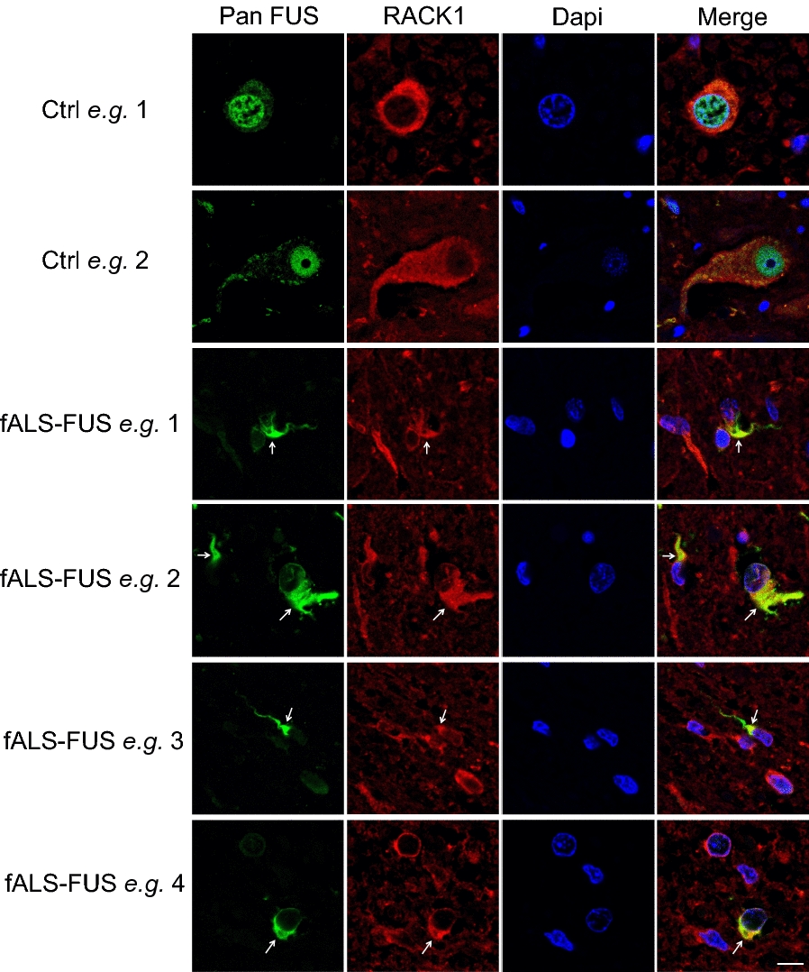
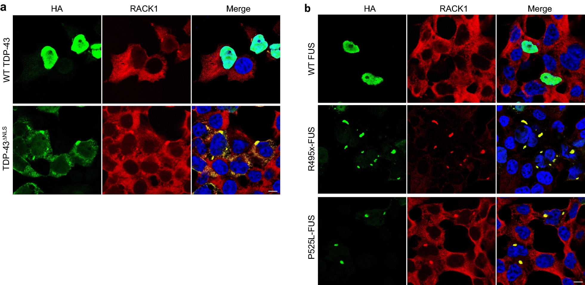
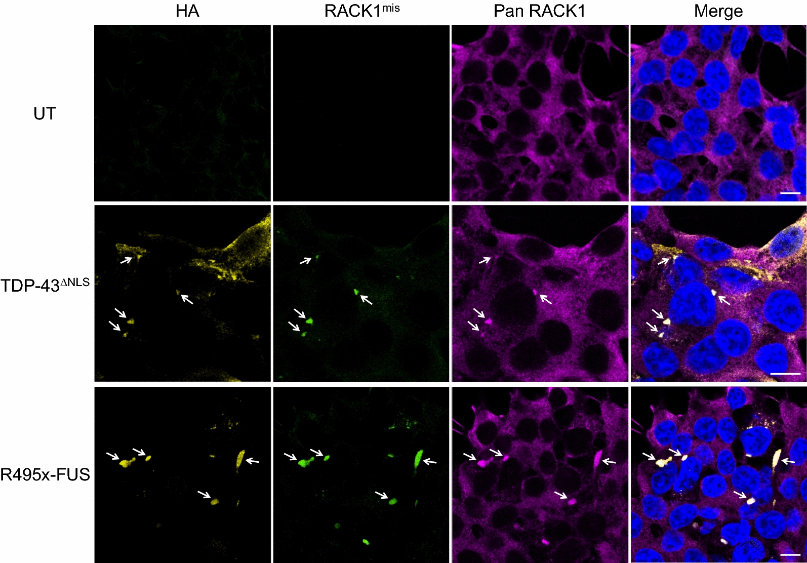
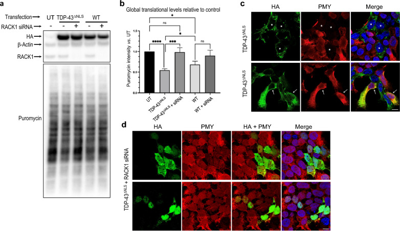
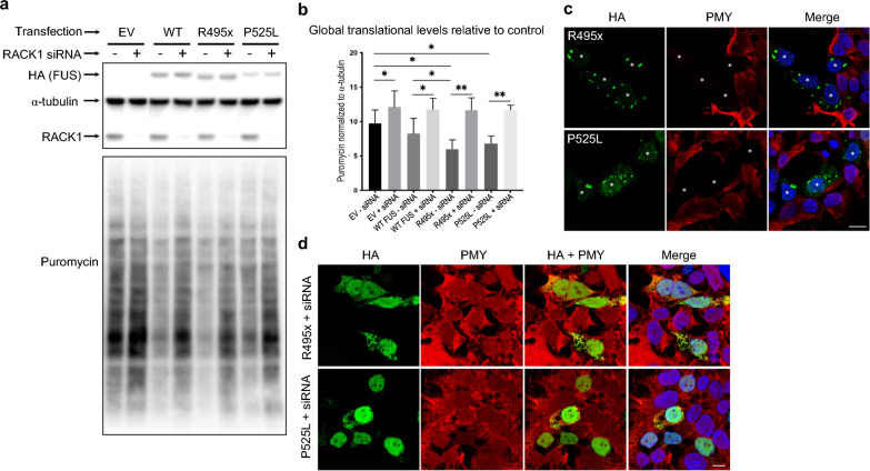
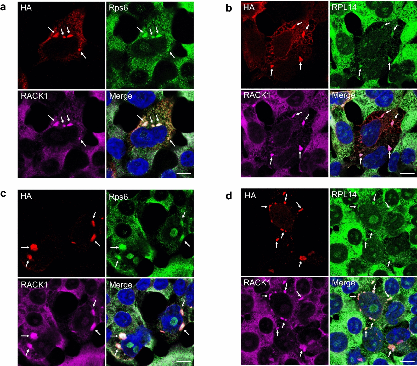
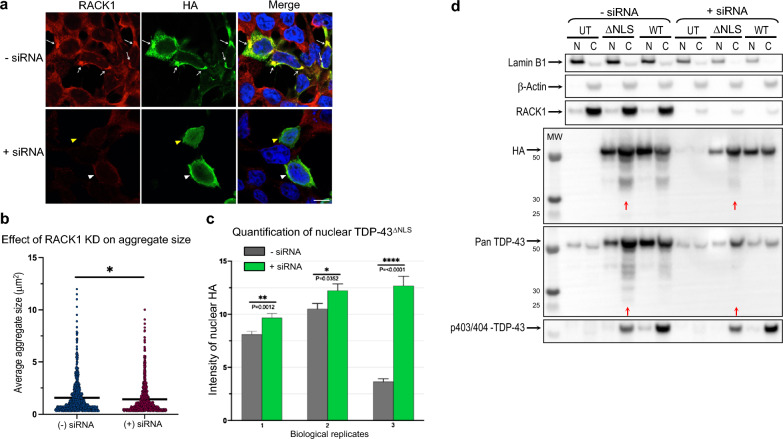
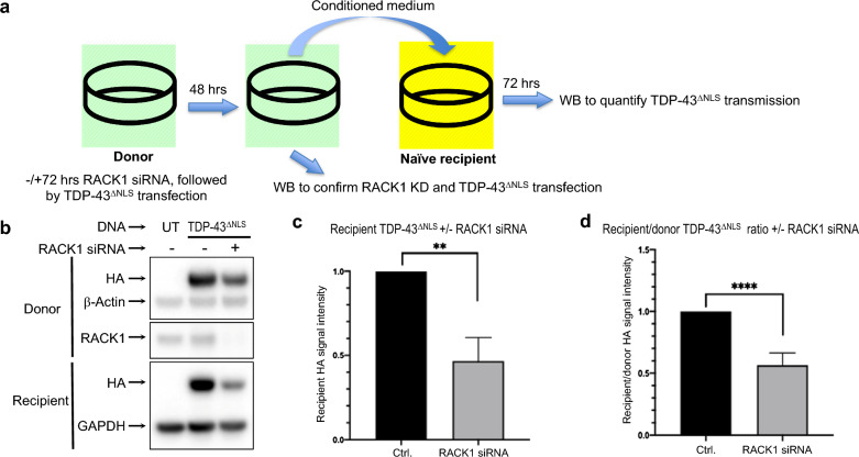
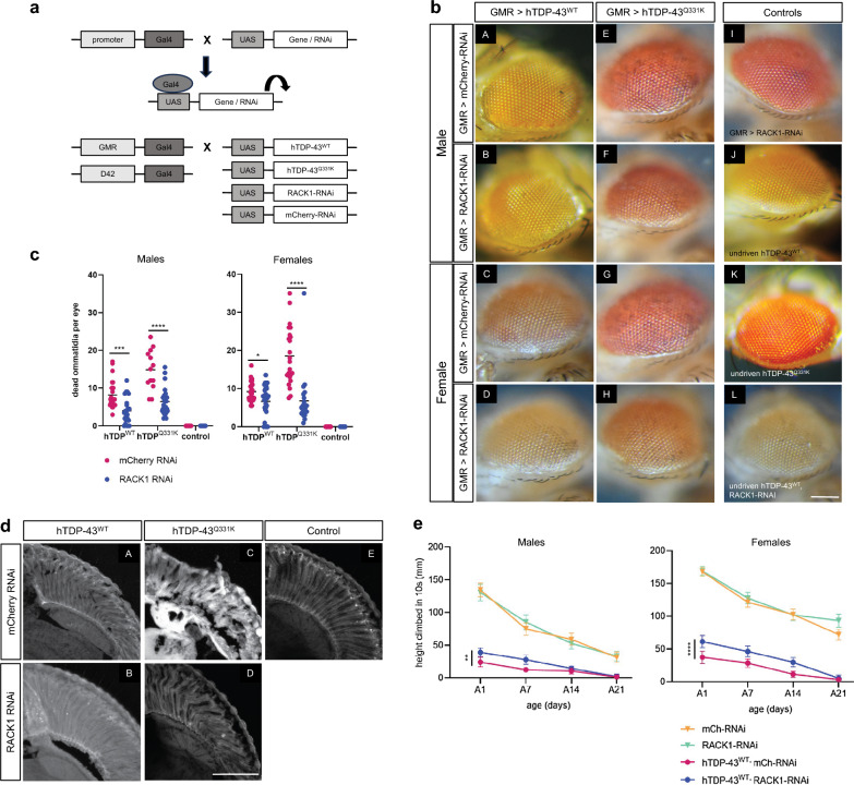
TAR DNA-binding protein 43 (TDP-43) and Fused in Sarcoma/Translocated in Sarcoma (FUS) are ribonucleoproteins associated with pathogenesis of amyotrophic lateral sclerosis (ALS) and frontotemporal lobar degeneration (FTLD). Under physiological conditions, TDP-43 and FUS are predominantly localized in the nucleus, where they participate in transcriptional regulation, RNA splicing and metabolism. In disease, however, they are typically mislocalized to the cytoplasm where they form aggregated inclusions. A number of shared cellular pathways have been identified that contribute to TDP-43 and FUS toxicity in neurodegeneration. In the present study, we report a novel pathogenic mechanism shared by these two proteins. We found that pathological FUS co-aggregates with a ribosomal protein, the Receptor for Activated C-Kinase 1 (RACK1), in the cytoplasm of spinal cord motor neurons of ALS, as previously reported for pathological TDP-43. In HEK293T cells transiently transfected with TDP-43 or FUS mutant lacking a functional nuclear localization signal (NLS; TDP-43ΔNLS and FUSΔNLS), cytoplasmic TDP-43 and FUS induced co-aggregation with endogenous RACK1. These co-aggregates sequestered the translational machinery through interaction with the polyribosome, accompanied by a significant reduction of global protein translation. RACK1 knockdown decreased cytoplasmic aggregation of TDP-43ΔNLS or FUSΔNLS and alleviated associated global translational suppression. Surprisingly, RACK1 knockdown also led to partial nuclear localization of TDP-43ΔNLS and FUSΔNLS in some transfected cells, despite the absence of NLS. In vivo, RACK1 knockdown alleviated retinal neuronal degeneration in transgenic Drosophila melanogaster expressing hTDP-43WT or hTDP-43Q331K and improved motor function of hTDP-43WT flies, with no observed adverse effects on neuronal health in control knockdown flies. In conclusion, our results revealed a novel shared mechanism of pathogenesis for misfolded aggregates of TDP-43 and FUS mediated by interference with protein translation in a RACK1-dependent manner. We provide proof-of-concept evidence for targeting RACK1 as a potential therapeutic approach for TDP-43 or FUS proteinopathy associated with ALS and FTLD.


Conflict of interest statement

N.R.C., J.M.K., and B.Z. are current employees of ProMIS Neurosciences. S.S.P. and S.C.C.H. have received consultation compensation from ProMIS Neurosciences. S.S.P. and B.Z. possess ProMIS stock options. N.R.C. and J.M.K. possess ProMIS shares and stock options. S.S.P. is the inventor on patent applications for Collective Coordinates computational modeling. N.R.C., S.S.P., J.M.K., and B.Z. are inventors on patent applications relating to conformation-specific epitopes in RACK1, antibodies thereto and methods related thereof.

Figures

Fig. 1
Fig. 1
Immunohistochemical analysis shows co-aggregation of RACK1 with FUS in the cytoplasm (arrows) in spinal cord sections of a fALS-FUS-R521C case, in contrast to normal nuclear expression of FUS in control (Ctrl). Nuclei stained with DAPI in merged images. Scale bar: 10 μm
Fig. 2
Fig. 2
Immunocytochemical analysis demonstrates expression of HA-tagged TDP-43ΔNLS (a, bottom row) or FUSΔNLS mutants, R495x- and P525L-FUS (b, middle & bottom rows), but not WT TDP-43 (a, top row) or WT-FUS (b, top row), which are primarily localized to the nucleus, is associated with co-aggregation of endogenous RACK1 in HEK293T cells. Nuclei stained with DAPI in merged images. Scale bars: 20 μm
Fig. 3
Fig. 3
Immunocytochemical analysis demonstrates reactivity of RACK1 misfolding specific antibody “RACK1mis” with RACK1 in cytoplasmic aggregates of HA-tagged TDP-43ΔNLS (middle row) or R495x-FUS (bottom row) transfected HEK293T cells but not with diffuse, non-aggregated RACK1 in the cytoplasm (stained by a Pan RACK1 antibody). RACK1mis shows no reactivity with endogenous, physiological RACK1 in un-transfected (UT, top row) cells. Nuclei stained with DAPI in merged images. Scale bars: 10 μm
Fig. 4
Fig. 4
RACK1 knockdown alleviates aggregation and global translational suppression by TDP-43ΔNLS and WT over-expression in HEK293T cells. a Representative SUnSET-Western Blot (WB) demonstrates that compared to control, expression of HA-tagged TDP-43ΔNLS and to a lesser extent over-expression of WT TDP-43, induces a significant reduction in puromycin (PMY) incorporation. b RACK1 siRNA KD alleviates global translational levels in both cases as determined by quantification of PMY band intensities normalized to loading control β-actin. c SUnSET-ICC shows inhibition of PMY incorporation by TDP-43ΔNLS (HA) expression preferably occurs in cells containing distinctive cytoplasmic aggregates (asterisks, top row). In cells where TDP-43ΔNLS displays a filamentary expression pattern (arrows, bottom row), PMY incorporation is comparable to neighbouring HA negative UT cells. d RACK1 KD alleviates aggregation, resulting in diffuse cytoplasmic expression (top row) or predominantly nuclear localization (bottom row) of TDP-43ΔNLS in a sub-population of transfected cells and correlating with normal PMY incorporation compared to neighbouring HA-negative UT cells. Nuclei stained with DAPI in merged images. Statistics: Ordinary one-way ANOVA Tukey multiple comparisons. n = 4 *p < 0.05; ***p < 0.001; ****p < 0.0001. Scale bars: 20 μm
Fig. 5
Fig. 5
RACK1 knockdown alleviates aggregation and restores global translational suppression by FUSΔNLS mutants. a Representative SUnSET-Western Blot (WB) demonstrates that compared to control empty vector (EV) transfected cells, expression of HA-tagged FUSΔNLS mutants, R495x- and P525L-FUS, and to a lesser extent over-expression of WT FUS, induces a significant reduction in puromycin (PMY) incorporation. b siRNA KD alleviates global translational levels in all cases as determined by quantification of PMY band intensities normalized to loading control α-tubulin. c SUnSET-ICC demonstrates HA-tagged FUSΔNLS mutants, R495x- and P525L-FUS, cause aggregation and completely inhibit PMY incorporation in transfected cells (asterisks). d RACK1 KD alleviates aggregation, resulting in diffuse cytoplasmic expression or predominantly nuclear localization of HA-tagged FUSΔNLS mutants, R495x- and P525L-FUS, in a sub-population of transfected cells and correlating with normal PMY incorporation compared to neighbouring HA-negative UT cells. Nuclei stained with DAPI in merged images. Statistics: Student’s t-test, unpaired, two-tailed. *p < 0.05 and **p < 0.005. n = 5 Error bars: SEM. Scale bars: 20 μm
Fig. 6
Fig. 6
Immunocytochemical analysis shows co-aggregation of HA-tagged TDP-43ΔNLS (a, b) or R495x-FUS (c, d) with RACK1 and eukaryotic 40S (Rps6) and 60S (RPL14) ribosome subunits (arrows). In neighbouring HA-negative UT cells, RACK1 is diffusely localized in the cytoplasm. Nuclei stained with DAPI in merged images. Scale bars: 10 μm
Fig. 7
Fig. 7
RACK1 knockdown not only reduces cytoplasmic aggregation but also increases nuclear localization of TDP-43ΔNLS in HEK293T cells. a Representative images showing that in contrast to control cells where distinctive aggregates of HA-tagged TDP-43ΔNLS are associated with RACK1 co-aggregation in the cytoplasm (arrows), RACK1 siRNA KD not only alleviates cytoplasmic aggregation (white arrowheads) but also leads to nuclear localization (yellow arrowheads) of TDP-43ΔNLS in a sub-population of transfected cells. Nuclei stained with DAPI in merged images. Scale bar: 10 μm. b Quantification of the average aggregate size in each individual TDP-43ΔNLS transfected cells shows a decrease following RACK1 KD. c Quantification of TDP-43ΔNLS nuclear localization from 3 biological repeats shows an increase as a result of RACK1 KD. d Western blot analysis confirms the quality of nucleocytoplasmic fractionation using nuclear and cytosolic protein markers, lamin B1 and β-Actin, respectively (top). TDP-43ΔNLS (HA) or total TDP-43 (pan TDP-43) from either fraction was normalized to β-actin or lamin B1. Quantification by densitometry shows that RACK1 KD decreases the cytoplasmic to nuclear ratio, fragmentation (red arrows, observed in 3 independent experiments), and phosphorylation of TDP-43ΔNLS (quantitated in one experiment). Statistics: Student’s t-test unpaired two-tailed. Error bars: SEM
Fig. 8
Fig. 8
RACK1 knockdown inhibits intercellular transmission of TDP-43ΔNLS in HEK293T cells. a Schematic illustration of the intercellular transmission procedure. Representative western blot (WB) b and densitometric quantification show reduced total HA-tagged TDP-43ΔNLS transmitted to naïve recipient cells c as well as a reduction in the recipient/donor ratio of TDP-43ΔNLS upon RACK1 KD. Statistics: Student’s t-test, unpaired two-tailed. **p < 0.005; ****p < 0.0001. n = 5
Fig. 9
Fig. 9
RACK1 knockdown significantly reduces hTDP-43-induced neuronal death and dysfunction in transgenic D. melanogaster. a Schematic illustration for the construction of transgenic D. melanogaster lines specifically targeting retinal (GMR-driven) or motor (D42-driven) neurons. b Representative images of fly eyes. Expression of hTDP-43WT (A–D) or hTDP-43Q331K (E–H) in retinal neurons cause neuronal death in both males and females, detected as darkened ommatidia. RACK1-RNAi reduces the appearance of dead ommatidia in all four conditions (B, D, F, H), compared to mCherry-RNAi control (A, C, E, G). Control flies with no dead ommatidia include those expressing RACK1-RNAi alone (I), and those harbouring unexpressed (undriven) transgenes (J–L). Scale bar: 250 mm. c Quantification of retinal neuronal death. Mann–Whitney test indicates a significant reduction in ommatidia death upon RACK1-RNAi KD in both males and females expressing either hTDP-43WT or hTDP-43Q331K. 21–33 flies were scored per condition. Statistics: Mann–Whitney test, *p < 0.05, ***p < 0.001. ****p < 0.0001. d Histological analysis of retina from flies expressing hTDP-43WT (A, B) or hTDP-43Q331K (C, D) in conjunction with control mCherry-RNAi (A, C) or RACK1-RNAi (B, D) show preservation of retinal architecture by RACK1 KD. Scale bar: 50 mm. e Both male and female hTDP-43WT transgenic flies show reduced climbing ability compared to mCherry-RNAi controls, which was significantly improved by RACK1-RNAi. Statistics: 2-way ANOVA. **p < 0.01, ****p < 0.0001. 60–100 flies were scored per condition. Error bars: SD

References

    1. Adams DR, Ron D, Kiely PA. RACK1, A multifaceted scaffolding protein: structure and function. Cell Commun Signal. 2011;9:22. doi: 10.1186/1478-811X-9-22. - DOI - PMC - PubMed
    1. Afroz T, Hock EM, Ernst P, Foglieni C, Jambeau M, Gilhespy LAB, Laferriere F, Maniecka Z, Pluckthun A, Mittl P, et al. Functional and dynamic polymerization of the ALS-linked protein TDP-43 antagonizes its pathologic aggregation. Nat Commun. 2017;8:45. doi: 10.1038/s41467-017-00062-0. - DOI - PMC - PubMed
    1. Arai T, Hasegawa M, Nonoka T, Kametani F, Yamashita M, Hosokawa M, Niizato K, Tsuchiya K, Kobayashi Z, Ikeda K, et al. Phosphorylated and cleaved TDP-43 in ALS, FTLD and other neurodegenerative disorders and in cellular models of TDP-43 proteinopathy. Neuropathology. 2010;30:170–181. doi: 10.1111/j.1440-1789.2009.01089.x. - DOI - PubMed
    1. Benarroch EE. Nucleocytoplasmic transport: Mechanisms and involvement in neurodegenerative disease. Neurology. 2019;92:757–764. doi: 10.1212/WNL.0000000000007305. - DOI - PubMed
    1. Birsa N, Bentham MP, Fratta P. Cytoplasmic functions of TDP-43 and FUS and their role in ALS. Semin Cell Dev Biol. 2020;99:193–201. doi: 10.1016/j.semcdb.2019.05.023. - DOI - PubMed

Publication types

MeSH terms

LinkOut - more resources