

Clinical Implementation and Evaluation of Auto-Segmentation Tools for Multi-Site Contouring in Radiotherapy
- PMID: 38111502
- PMCID: PMC10726238
- DOI: 10.1016/j.phro.2023.100515
Clinical Implementation and Evaluation of Auto-Segmentation Tools for Multi-Site Contouring in Radiotherapy
Abstract
Background and purpose: Tools for auto-segmentation in radiotherapy are widely available, but guidelines for clinical implementation are missing. The goal was to develop a workflow for performance evaluation of three commercial auto-segmentation tools to select one candidate for clinical implementation.
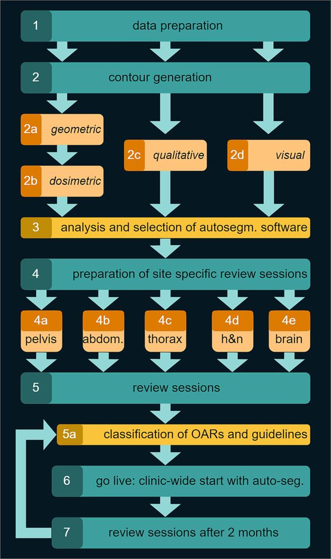
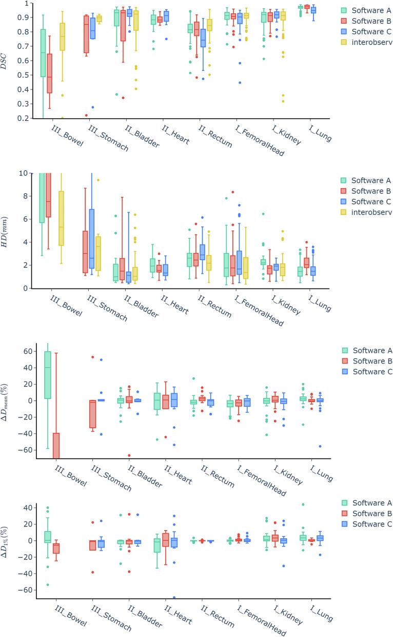
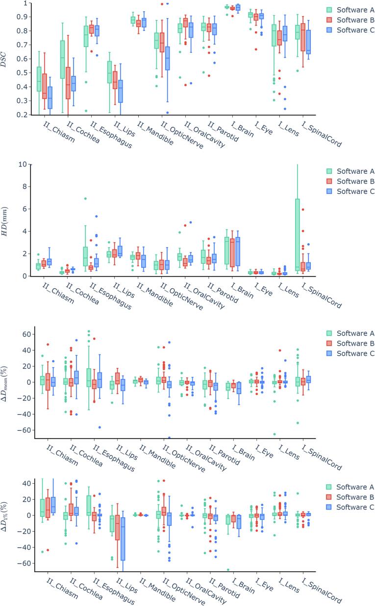
Materials and methods: One hundred patients with six treatment sites (brain, head-and-neck, thorax, abdomen, and pelvis) were included. Three sets of AI-based contours for organs-at-risk (OAR) generated by three software tools and manually drawn expert contours were blindly rated for contouring accuracy. The dice similarity coefficient (DSC), the Hausdorff distance, and a dose/volume evaluation based on the recalculation of the original treatment plan were assessed. Statistically significant differences were tested using the Kruskal-Wallis test and the post-hoc Dunn Test with Bonferroni correction.
Results: The mean DSC scores compared to expert contours for all OARs combined were 0.80 ± 0.10, 0.75 ± 0.10, and 0.74 ± 0.11 for the three software tools. Physicians' rating identified equivalent or superior performance of some AI-based contours in head (eye, lens, optic nerve, brain, chiasm), thorax (e.g., heart and lungs), and pelvis and abdomen (e.g., kidney, femoral head) compared to manual contours. For some OARs, the AI models provided results requiring only minor corrections. Bowel-bag and stomach were not fit for direct use. During the interdisciplinary discussion, the physicians' rating was considered the most relevant.
Conclusion: A comprehensive method for evaluation and clinical implementation of commercially available auto-segmentation software was developed. The in-depth analysis yielded clear instructions for clinical use within the radiotherapy department.
Keywords: Auto-segmentation; Deep Learning; Radiotherapy; Segmentation.
© 2023 The Author(s).
Conflict of interest statement
The authors declare that they have no known competing financial interests or personal relationships that could have appeared to influence the work reported in this paper.
Figures
References
LinkOut - more resources
Full Text Sources
