

Optical coherence tomography angiography for macular microvessels in ischemic branch retinal vein occlusion treated with conbercept: predictive factors for the prognosis
- PMID: 38111937
- PMCID: PMC10700074
- DOI: 10.18240/ijo.2023.12.18
Optical coherence tomography angiography for macular microvessels in ischemic branch retinal vein occlusion treated with conbercept: predictive factors for the prognosis
Abstract
Aim: To evaluate the predicative factors of visual prognosis using optical coherence tomography angiography (OCTA) in ischemic branch retinal vein occlusion (BRVO) patients with macular edema (ME) after anti-vascular endothelial growth factor (VEGF) treatment.
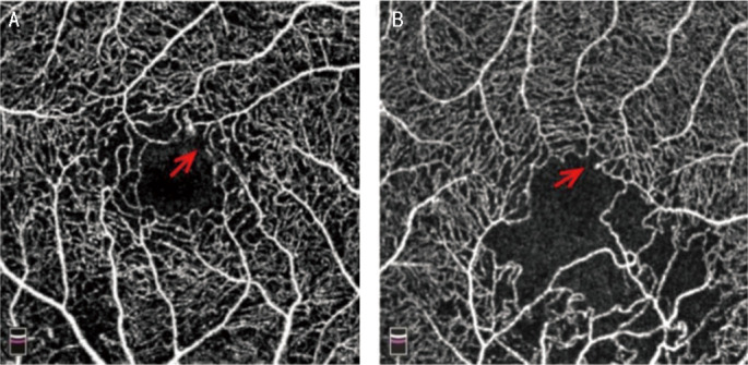
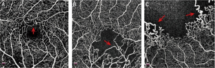
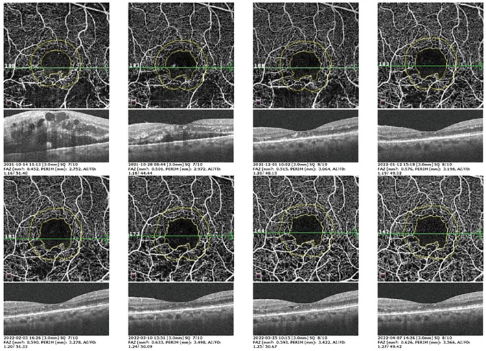
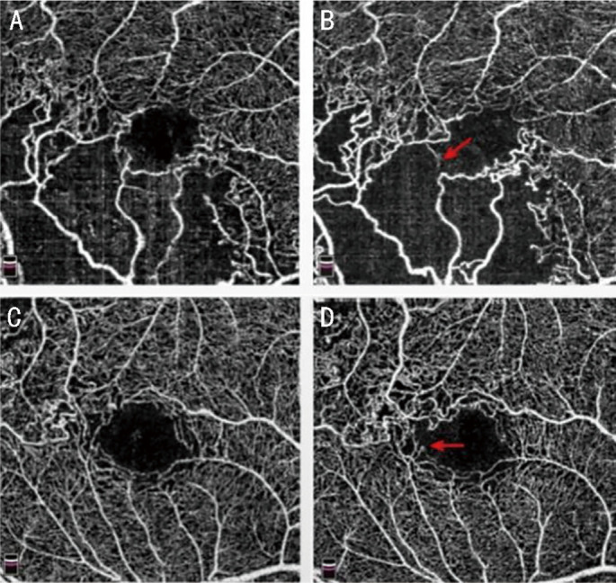
Methods: In this retrospective analysis, data from 60 patients (60 eyes) with a definite diagnosis of ischemic BRVO with ME by fundus fluorescein angiography (FFA) were studied. The eyes with ME according to spectral domain optical coherence tomography (SD-OCT) underwent intravitreal conbercept (IVC) and 3+pro re nata (PRN) regimen. The injection times were recorded. Two weeks after injection, fundus laser photocoagulation was performed in the non-perfusion area of the retina. The patients were followed up once a month for 6mo. The best-corrected visual acuity (BCVA), foveal avascular zone (FAZ), and A-circularity index (AI), at 6mo and the baseline were compared.
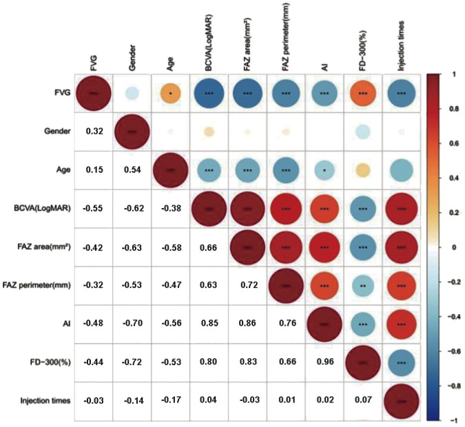
Results: All patients showed significant improvement in BCVA from 0.82±0.32 to 0.39±0.11 logMAR (P<0.001). The mean central macular thickness (CMT) significantly decreased from 476.22±163.54 to 298.66±109.23 µm. Both the FAZ area and AI at 6mo were significantly higher than those at the baseline: the FAZ area increased (0.38±0.02 vs 0.39±0.02 mm2, P<0.05); the AI increased (1.27±0.02 vs 1.31±0.01, P=0.000). The baseline BCVA showed a significantly positive correlation with the baseline FAZ area, FAZ perimeter (PERIM) and AI, final visual gain (FVG) and injection times, respectively (P<0.001). FVG showed a significantly negative correlation with the FAZ area, PERIM, AI and injection times, but a significantly positive correlation with vessel densities (VDs) 300 µm area around FAZ (FD-300; P<0.001). Injection times was positively correlated with the baseline FAZ area, and AI, but inversely correlated with the baseline FD-300 (P<0.001). However macular ischemia was noted in 5 cases during follow-up.
Conclusion: Using OCTA to observe macular ischemia and quantify parameters can better predict the final visual prognosis of patients before treatment. The changes in FAZ parameters may influence the visual prognosis and injection times.
Keywords: branch retinal vein occlusion; conbercept; foveal avascular zone; macular edema; optical coherence tomography angiography.
International Journal of Ophthalmology Press.
Figures

Similar articles
-
Quantitative Analysis of Retinal Microvascular Changes after Conbercept Therapy in Branch Retinal Vein Occlusion Using Optical Coherence Tomography Angiography.Ophthalmologica. 2019;242(2):69-80. doi: 10.1159/000499608. Epub 2019 May 21. Ophthalmologica. 2019. PMID: 31112969
-
Conbercept improves macular microcirculation and retinal blood supply in the treatment of nonischemic branch retinal vein occlusion macular edema.J Clin Lab Anal. 2022 Dec;36(12):e24774. doi: 10.1002/jcla.24774. Epub 2022 Nov 21. J Clin Lab Anal. 2022. PMID: 36408725 Free PMC article.
-
Microvascular changes after conbercept therapy in central retinal vein occlusion analyzed by optical coherence tomography angiography.Int J Ophthalmol. 2019 May 18;12(5):802-808. doi: 10.18240/ijo.2019.05.16. eCollection 2019. Int J Ophthalmol. 2019. PMID: 31131240 Free PMC article.
-
Quantification of retinal microvasculature and neurodegeneration changes in branch retinal vein occlusion after resolution of cystoid macular edema on optical coherence tomography angiography.Indian J Ophthalmol. 2019 Nov;67(11):1864-1869. doi: 10.4103/ijo.IJO_1554_18. Indian J Ophthalmol. 2019. PMID: 31638051 Free PMC article.
-
Changes in Macular Microvascular Structure in Macular Edema Secondary to Branch Retinal Vein Occlusion Treated with Antivascular Endothelial Growth Factor for One Year.J Ophthalmol. 2021 May 17;2021:6645452. doi: 10.1155/2021/6645452. eCollection 2021. J Ophthalmol. 2021. PMID: 34055397 Free PMC article.
Cited by
-
Efficacy of Ozurdex implants as second-line therapy for non-responders to anti-VEGF in retinal vein occlusion-associated macular edema: a retrospective cohort study.Ir J Med Sci. 2025 Apr;194(2):745-750. doi: 10.1007/s11845-025-03881-z. Epub 2025 Jan 24. Ir J Med Sci. 2025. PMID: 39853533 Free PMC article.
References
-
- Rogers SL, McIntosh RL, Lim L, Mitchell P, Cheung N, Kowalski JW, Nguyen HP, Wang JJ, Wong TY. Natural history of branch retinal vein occlusion: an evidence-based systematic review. Ophthalmology. 2010;117(6):1094–1101.e5. - PubMed
-
- Jaulim A, Ahmed B, Khanam T, Chatziralli IP. Branch retinal vein occlusion: epidemiology, pathogenesis, risk factors, clinical features, diagnosis, and complications. An update of the literature. Retina. 2013;33(5):901–910. - PubMed
-
- Sangroongruangsri S, Ratanapakorn T, Wu O, Anothaisintawee T, Chaikledkaew U. Comparative efficacy of bevacizumab, ranibizumab, and aflibercept for treatment of macular edema secondary to retinal vein occlusion: a systematic review and network meta-analysis. Expert Rev Clin Pharmacol. 2018;11(9):903–916. - PubMed
-
- Spaide RF, Klancnik JM, Jr, Cooney MJ. Retinal vascular layers imaged by fluorescein angiography and optical coherence tomography angiography. JAMA Ophthalmol. 2015;133(1):45. - PubMed
LinkOut - more resources
Full Text Sources