. 2024 May;48(5):514-524.
doi: 10.1111/aor.14698. Epub 2023 Dec 19.

Investigation of the role of von Willebrand factor in shear-induced platelet activation and functional alteration under high non-physiological shear stress

Affiliations

Investigation of the role of von Willebrand factor in shear-induced platelet activation and functional alteration under high non-physiological shear stress

Dong Han et al. Artif Organs. 2024 May.

Abstract

Background: von Willebrand factor (vWF) plays a crucial role in physiological hemostasis through platelet and subendothelial collagen adhesion. However, its role in shear-induced platelet activation and functional alteration under non-physiological conditions common to blood-contacting medical devices (BCMDs) is not well investigated.

Methods: Fresh healthy human blood was treated with an anti-vWF antibody to block vWF-GPIbα interaction. Untreated blood was used as a control. They were exposed to three levels of non-physiological shear stress (NPSS) (75, 125, and 175 Pa) through a shearing device with an exposure time of 0.5 s to mimic typical shear conditions in BCMDs. Flow cytometric assays were used to measure the expression levels of PAC-1 and P-Selectin and platelet aggregates for platelet activation and the expression levels of GPIbα, GPIIb/IIIa, and GPVI for receptor shedding. Collagen/ristocetin-induced platelet aggregation capacity was characterized by aggregometry.

Results: The levels of platelet activation and aggregates increased with increasing NPSS in the untreated blood. More receptors were lost with increasing NPSS, resulting in a decreased capacity of collagen/ristocetin-induced platelet aggregation. In contrast, the increase in platelet activation and aggregates after exposure to NPSS, even at the highest level of NPSS, was significantly lower in treated blood. Nevertheless, there was no notable difference in receptor shedding, especially for GPIIb/IIIa and GPVI, between the two blood groups at the same level of NPSS. The block of vWF exacerbated the decreased capacity of collagen/ristocetin-induced platelet aggregation.

Conclusions: High NPSS activates platelets mainly by enhancing the vWF-GPIbα interaction. Platelet activation and receptor shedding induced by high NPSS likely occur through different pathways.

Keywords: blood‐contacting medical devices; non‐physiological shear stress; platelet activation; platelet dysfunction; von Willebrand factor (vWF).


Figures

Figure 1.
Figure 1.
Schematic cross-section view of the blood-shearing device. Blood is sheared in the narrow gap between the levitated rotating rotor and static housing where Couette flow is generated. The level of shear stress is controlled by the rotating speed of the rotor and the exposure time is controlled by the flow rate at the blood inlet pushed a syringe pump. Sheared blood sample is collected from sample port, and waste port allows excessive blood to exit.
Figure 2.
Figure 2.
A typical image of the distribution of vWF multimers in plasma in anti-vWF antibody treated and untreated samples, both at baseline and at three shear conditions.
Figure 3.
Figure 3.
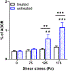
The surface expression and mean fluorescent intensity (MFI) of PAC-1 on platelets from the anti-vWF antibody-treated and untreated blood samples after NPSS exposure. (a) Percentage change of activated platelets indicated by PAC-1 surface expression. (c) MFI of activated platelets indicated by PAC-1 surface expression. (# represents the significant difference between its corresponding baseline sample, #p < 0.05, ###p < 0.001, n = 6; * represents significant difference between two groups under the same level of NPSS, **p < 0.01, ***p < 0.001, n = 6).
Figure 4.
Figure 4.
The surface expression and mean fluorescent intensity (MFI) of CD62P+ on platelets from the anti-vWF antibody-treated and untreated blood after NPSS exposure. (a) Percentage change of activated platelets indicated by CD62P+. (c) MFI of activated platelets indicated by CD62P+ surface expression. (# represents the significant difference between its corresponding baseline sample, #p < 0.05, ###p < 0.001, n = 6; * represents significant difference between two groups under the same level of NPSS, *p < 0.05, **p < 0.01, ***p < 0.001, n = 6).
Figure 5.
Figure 5.
The quantification of the platelet GP receptor expression from anti-vWF antibody-treated and untreated blood after NPSS exposure. (a) Fold change of GPIbα MFI in the baseline and sheared blood samples. (b) Fold change of GPIIb/IIIa MFI in the baseline and sheared blood samples. (c) Fold change of GPVI MFI in the baseline and sheared blood samples. (# represents the significant difference between its corresponding baseline sample, #p < 0.05, ###p < 0.001, n = 6).
Figure 6.
Figure 6.
Percentage change of platelet aggregates in anti-vWF antibody-treated and untreated blood after NPSS exposure. (# represents the significant difference between its corresponding baseline sample, ##p < 0.01, ###p < 0.001, n = 6; * represents significant difference between two groups under the same level of NPSS, **p < 0.01, ***p < 0.001, n = 6).
Figure 7.
Figure 7.
Patterns of ristocetin/collagen-induced platelet aggregation in anti-vWF antibody-treated and untreated blood after NPSS exposure. (a) Ristocetin-induced platelet aggregation for anti-vWF antibody-untreated blood. (b) Ristocetin-induced platelet aggregation for anti-vWF antibody-treated blood. (c) Collagen-induced platelet aggregation for anti-vWF antibody-untreated blood. (d) Collagen-induced platelet aggregation for anti-vWF antibody-treated blood.
Figure 8.
Figure 8.
Quantification of platelet aggregation capability change (induced by collagen and ristocetin) in anti-vWF antibody-treated and untreated blood samples after NPSS exposure. The duration of the platelet aggregation curve changing was recorded for 6 min, and the platelet aggregation is indicated by AUC. (a) Change of platelet aggregation ability induced by ristocetin. (b) Fold change of platelet aggregation ability induced by collagen. (# represents the significant difference between its corresponding baseline sample, #p < 0.05, ###p < 0.001, n = 6; * represents significant difference between two groups under the same level of NPSS, ***p < 0.001, n = 6).

References

    1. Mazzeffi M, Greenwood J, Tanaka K, Menaker J, Rector R, Herr D, et al. Bleeding, Transfusion, and Mortality on Extracorporeal Life Support: ECLS Working Group on Thrombosis and Hemostasis. In: Annals of Thoracic Surgery. Elsevier; USA; 2016. p. 682–9. - PubMed
    1. Shah P, Tantry US, Bliden KP, Gurbel PA. Bleeding and thrombosis associated with ventricular assist device therapy. Journal of Heart and Lung Transplantation. 2017;36:1164–73. - PubMed
    1. Starling RC, Naka Y, Boyle AJ, Gonzalez-Stawinski G, John R, Jorde U, et al. Results of the post-U.S. food and drug administration-approval study with a continuous flow left ventricular assist device as a bridge to heart transplantation: A prospective study using the INTERMACS (Interagency Registry for Mechanically Assisted Circulatory Support). J Am Coll Cardiol. 2011;57:1890–8. - PubMed
    1. Gupta P, McDonald R, Chipman CW, Stroud M, Gossett JM, Imamura M, et al. 20-year experience of prolonged extracorporeal membrane oxygenation in critically III children with cardiac or pulmonary failure. Annals of Thoracic Surgery. 2012;93:1584–90. - PubMed
    1. Chen Z, Zhang J, Kareem K, Tran D, Conway RG, Arias K, et al. Device-induced platelet dysfunction in mechanically assisted circulation increases the risks of thrombosis and bleeding. Artif Organs. 2019;43:745–55. - PMC - PubMed

LinkOut - more resources