. 2024 Jan 24;90(1):e0154823.
doi: 10.1128/aem.01548-23. Epub 2023 Dec 19.

FlhF affects the subcellular clustering of WspR through HsbR in Pseudomonas aeruginosa

Affiliations

FlhF affects the subcellular clustering of WspR through HsbR in Pseudomonas aeruginosa

Congcong Guan et al. Appl Environ Microbiol. .

Abstract

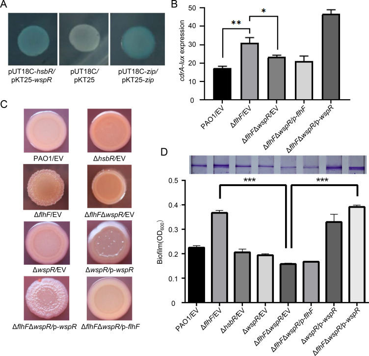
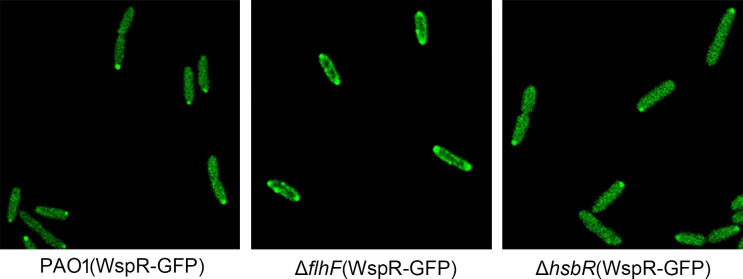
In bacteria, the second messenger cyclic di-GMP (c-di-GMP) is synthesized and degraded by multiple diguanylate cyclases (DGCs) and phosphodiesterases. A high level of c-di-GMP induces biofilm formation and represses motility. WspR, a hybrid response regulator DGC, produces c-di-GMP when it is phosphorylated. FlhF, a signal recognition particle-type GTPase, is initially localized to the cell poles and is indispensable for polar flagellar localization in Pseudomonas aeruginosa. In this study, we report that deletion of flhF affected biofilm formation and the c-di-GMP level in P. aeruginosa. Phenotypic analysis of a flhF knockout mutant revealed increased biofilm formation, wrinkled colonies on Congo red agar, and an elevated c-di-GMP level compared to the wild-type strain, PAO1. Yeast and bacterial two-hybrid systems showed that FlhF binds to the response regulator HsbR, and HsbR binds to WspR. Deletion of hsbR or wspR in the ΔflhF background abolished the phenotype of ΔflhF. In addition, confocal microscopy demonstrated that WspR-GFP was distributed throughout the cytoplasm and formed a visible cluster at one cell pole in PAO1 and ΔhsbR, but it was mainly distributed as visible clusters at the lateral side of the periplasm and with visible clusters at both cell poles in ΔflhF. These findings suggest that FlhF influences the subcellular cluster and localization of WspR and negatively modulates WspR DGC activity in a manner dependent on HsbR. Together, our findings demonstrate a novel mechanism for FlhF modulating the lifestyle transition between motility and biofilm via HsbR to regulate the DGC activity of WspR.IMPORTANCECyclic di-GMP (c-di-GMP) is a second messenger that controls flagellum biosynthesis, adhesion, virulence, motility, exopolysaccharide production, and biofilm formation in bacteria. Recent research has shown that distinct diguanylate cyclases (DGCs) or phosphodiesterases (PDEs) produce highly specific outputs. Some DGCs and PDEs contribute to the total global c-di-GMP concentration, but others only affect local c-di-GMP in a microenvironment. However, the underlying mechanisms are unclear. Here, we report that FlhF affects the localization and DGC activity of WspR via HsbR and is implicated in local c-di-GMP signaling in Pseudomonas aeruginosa. This study establishes the link between the c-di-GMP signaling system and the flagellar localization and provides insight for understanding the complex regulatory network of c-di-GMP signaling.

Keywords: FlhF; WspR; biofilm formation; c-di-GMP; subcellular clustering.


Conflict of interest statement

The authors declare no conflict of interest.

Figures

Fig 1
Fig 1
The ΔflhF mutant displays high c-di-GMP phenotypes. (A) The ΔflhF mutant is defective for swarming and swimming motility. (B) Deletion of flhF increased the biofilm formation in P. aeruginosa. Biofilm formation by the indicated strains was displayed with crystal violet staining (top) and quantified by optical density measurement (bottom). (C) Congo red binding phenotype was visualized after 2 days of incubation. The wrinkly phenotype was observed in the indicated P. aeruginosa strains. (D) The relative intracellular level of c-di-GMP was measured with the transcriptional pKD-cdrA reporter. (E) Western blotting analysis of CdrA-Flag showed the relative level of c-di-GMP in bacterial strains. PAO1 and ΔflhF mutant harbored a control plasmid (pAK1900). The plasmid p-2133 was used to express the phosphodiesterase PA2133. The protein level of CdrA-Flag from the indicated strains was examined by western blotting. The tagged proteins were detected using a Flag antibody. RNA polymerase α (α-RNAP) antibody was used as a loading control. EV represents the empty vector in this and subsequent experiments. Data are shown as mean ± SEM from three experiments at least. The error bars indicate standard deviations (*P < 0.05, **P < 0.01, and ***P < 0.001, Student’s t-test).
Fig 2
Fig 2
FlhF regulates biofilm formation via interaction with HsbR. (A) A yeast two-hybrid assay reveals an interaction between FlhF and HsbR. The yeast two-hybrid reporter strain AH109 containing the indicated plasmids was incubated on SD/-Trp/-Leu medium and SD/-Trp/-Leu/-His/-Ade medium at 30°C for 2 days. pGADT7-largeT and pGBKT7-p53 were used as positive controls; pGADT7 and pGBKT7 were used as negative controls. The plasmids harbored by the AH109 strains are indicated on the right. (B) Colony morphologies were demonstrated by growth on Congo red agar plates after 2 days of incubation. The wrinkly phenotype was observed in the indicated P. aeruginosa strains. (C) Biofilm formation by the indicated strains was displayed with crystal violet staining (top) and quantified by optical density measurement (bottom). Each experiment was repeated three times at least. The error bars indicate standard deviations (**P < 0.01 and ****P < 0.0001, Student’s t-test).
Fig 3
Fig 3
Analyses of interactions between FlhF and HsbR subdomains. (A) Schematic representation of the HsbR subdomains. REC domain (amino acids 13–125), PP2C domain (amino acids 186–386), and HATP domain (amino acids 442–571). (B) Yeast two-hybrid analyses of interactions between FlhF and HsbR subdomains. The reporter strain AH109 containing the indicated plasmids was incubated on SD/-Trp/-Leu medium and SD/-Trp/-Leu/-His/-Ade medium at 30°C for 2 days. pGADT7-largeT and pGBKT7-p53 were used as positive controls; pGADT7 and pGBKT7 were used as negative controls. The plasmids harbored by the AH109 strains are indicated on the right. Each experiment was repeated three times at least.
Fig 4
Fig 4
HsbA and HsbD are not involved in the phenotypes of ΔflhF. Biofilm formation by the indicated P. aeruginosa strains was displayed with crystal violet staining (top), and the wrinkly phenotype of indicated strains was observed by growth on Congo red agar plates after 2 days of incubation (bottom). Each experiment was repeated three times at least.
Fig 5
Fig 5
WspR impacts the phenotypes of ΔflhF. (A) The bacterial two-hybrid assay reveals an interaction between HsbR and WspR. The bacterial two-hybrid reporter Escherichia coli BTH101 recombinant strains harboring the indicated plasmids were separately streaked on LB/X-Gal/IPTG (isopropyl β-D-thiogalactoside) plates. The pUT18C-zip and pKT25-zip plasmids were used as positive controls. The pUT18C and pKT25 plasmids were used as negative controls. (B) The relative intracellular level of c-di-GMP was measured with the transcriptional pKD-cdrA reporter. (C) Colony morphologies were demonstrated by growth on Congo red agar plates after 2 days of incubation. The wrinkly phenotype was observed in the indicated P. aeruginosa strains. (D) Biofilm formation by the indicated strains was displayed with crystal violet staining (top) and quantified by optical density measurement (bottom). Each experiment was repeated three times at least. The error bars indicate standard deviations (*P < 0.05, **P < 0.01, and ***P < 0.001, Student’s t-test).
Fig 6
Fig 6
Fluorescence micrographs of strain PAO1 derivatives expressing the WspR-GFP. Overnight cultures of wild-type PAO1, ΔflhF mutant, and the ΔhsbR expressing WspR-GFP were subcultured in fresh Luria–Bertani (LB) with 0.5 mM IPTG for 3 h. Bacterial cells were fixed and examined by fluorescence microscopy.
Fig 7
Fig 7
Model for FlhF affecting the sublocation of WspR through HsbR and yielding a local c-di-GMP. Green gradient in the cell illustrates the distribution of c-di-GMP levels. (A) Model for the FlhF-modulated c-di-GMP production through HsbR and WspR. FlhF inhibits the DGC activity of WspR by inhibiting the HsbR-mediated phosphorylation of WspR. WspR-P tends to form visible subcellular clusters with high DGC activity. (B) WspR-GFP was diffused in the cytoplasm and formed a visible cluster at one pole in PAO1, yielding low concentration c-di-GMP and inducing motility. WspR could produce a local high c-di-GMP concentration at the pole where it formed clusters. (C) WspR formed oligomers and localized in the periphery of the ΔflhF cell, yielding an increased global concentration of c-di-GMP, resulting in biofilm formation.

Similar articles

Cited by

References

    1. Mulcahy LR, Isabella VM, Lewis K. 2014. Pseudomonas aeruginosa biofilms in disease. Microb Ecol 68:1–12. doi:10.1007/s00248-013-0297-x - DOI - PMC - PubMed
    1. Lyczak JB, Cannon CL, Pier GB. 2002. Lung infections associated with cystic fibrosis. Clin Microbiol Rev 15:194–222. doi:10.1128/CMR.15.2.194-222.2002 - DOI - PMC - PubMed
    1. Høiby N, Bjarnsholt T, Moser C, Bassi GL, Coenye T, Donelli G, Hall-Stoodley L, Holá V, Imbert C, Kirketerp-Møller K, Lebeaux D, Oliver A, Ullmann AJ, Williams C, ESCMID Study Group for Biofilms and Consulting External Expert Werner Zimmerli . 2015. ESCMID guideline for the diagnosis and treatment of biofilm infections 2014. Clin Microbiol Infect 21 Suppl 1:S1–25. doi:10.1016/j.cmi.2014.10.024 - DOI - PubMed
    1. Sauer K, Camper AK, Ehrlich GD, Costerton JW, Davies DG. 2002. Pseudomonas aeruginosa displays multiple phenotypes during development as a biofilm. J Bacteriol 184:1140–1154. doi:10.1128/jb.184.4.1140-1154.2002 - DOI - PMC - PubMed
    1. Sauer Karin, Stoodley P, Goeres DM, Hall-Stoodley L, Burmølle M, Stewart PS, Bjarnsholt T. 2022. The biofilm life cycle: expanding the conceptual model of biofilm formation. Nat Rev Microbiol 20:608–620. doi:10.1038/s41579-022-00767-0 - DOI - PMC - PubMed

Publication types

MeSH terms

LinkOut - more resources