. 2024 Jan 16;15(1):e0203623.
doi: 10.1128/mbio.02036-23. Epub 2023 Dec 19.

Requirement of GrgA for Chlamydia infectious progeny production, optimal growth, and efficient plasmid maintenance

Affiliations

Requirement of GrgA for Chlamydia infectious progeny production, optimal growth, and efficient plasmid maintenance

Bin Lu et al. mBio. .

Abstract

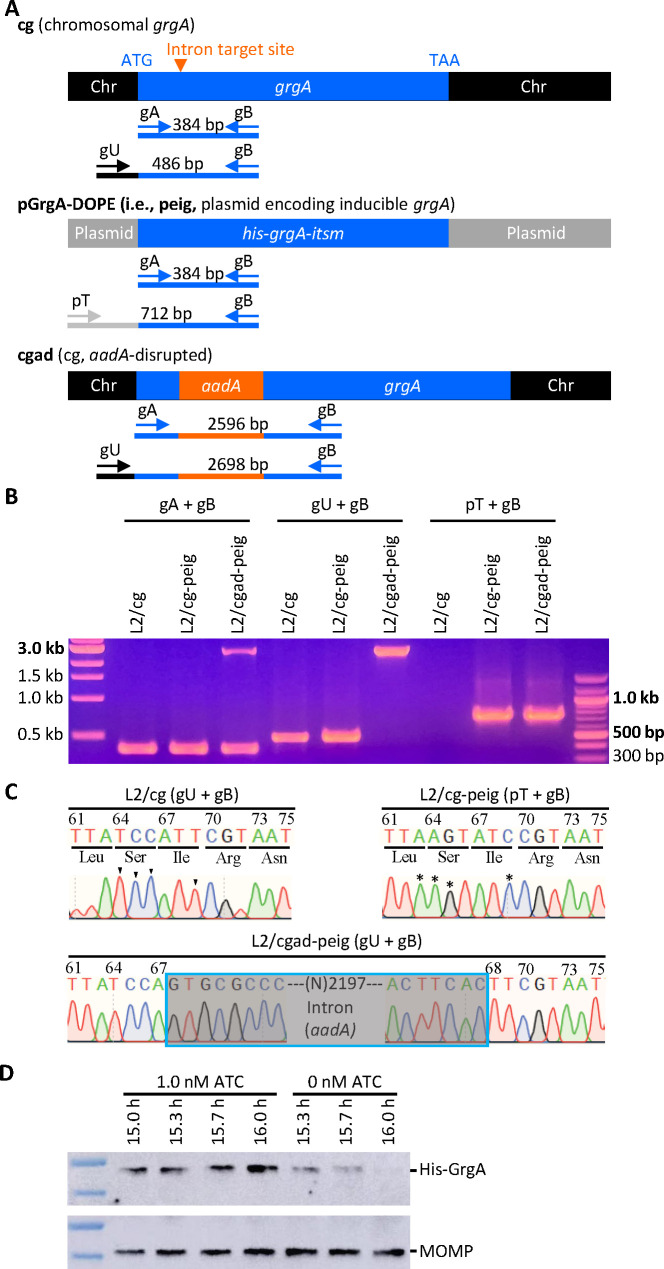
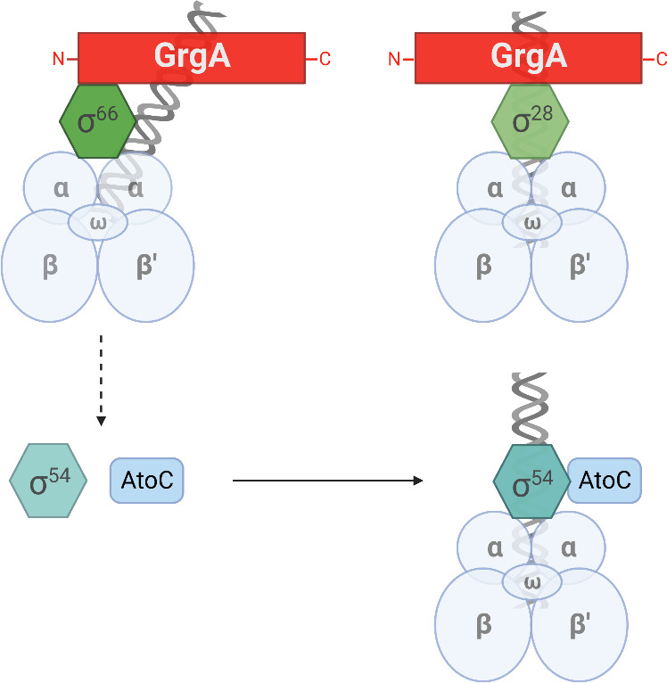
Hallmarks of the developmental cycle of the obligate intracellular pathogenic bacterium Chlamydia are the primary differentiation of the infectious elementary body (EB) into the proliferative reticulate body (RB) and the secondary differentiation of RBs back into EBs. The mechanisms regulating these transitions remain unclear. In this report, we developed an effective novel strategy termed dependence on plasmid-mediated expression (DOPE) that allows for the knockdown of essential genes in Chlamydia. We demonstrate that GrgA, a Chlamydia-specific transcription factor, is essential for the secondary differentiation and optimal growth of RBs. We also show that GrgA, a chromosome-encoded regulatory protein, controls the maintenance of the chlamydial virulence plasmid. Transcriptomic analysis further indicates that GrgA functions as a critical regulator of all three sigma factors that recognize different promoter sets at developmental stages. The DOPE strategy outlined here should provide a valuable tool for future studies examining chlamydial growth, development, and pathogenicity.

Keywords: Chlamydia; GrgA; regulation of gene expression; transcription factors; transcriptional regulation.


Conflict of interest statement

The authors declare no conflict of interest.

Figures

Fig 1
Fig 1
Confirmation of the disruption of the chromosome-encoded grgA by group II intron and re-expression of GrgA from a transformed plasmid in the DOPE system. (A) Schematic drawings of grgA alleles, locations of intron target site, diagnostic primers, and sizes of PCR products obtained with different sets of primers. Abbreviations: itsm, intron target site mutated; Chr, chromosome. (B) Gel image of PCR products amplified with DNA of wild-type C. trachomatis L2 with intact chromosomal grgA (L2/cg), L2/cg transformed with the his-grgA-itsm expression plasmid pGrgA-DOPE (L2/cg-peig), and L2 with aadA-disrupted chromosomal grgA complemented with pGrgA-DOPE (L2/cgad-peig) using primer sets shown in (A). (C) Sanger sequencing tracings of PCR products showing the intron target site in L2/cg, mutations surrounding this site conferring resistance to intron targeting in peig, and grgA-intron joint regions in the chromosome of L2/cg-peig. Wild-type bases and corresponding mutated bases are shown with arrowheads and asterisks, respectively. (D) Western blotting showing time-dependent loss of His-GrgA in L2/cgad-peig upon ATC withdrawal. HeLa cells infected with L2/cgad-peig were cultured in the presence of 1 nM ATC. Three cultures were switched to ATC-free medium at 15 h postinoculation hpi. Cultures were harvested with SDS-PAGE sample buffer at indicated times and resolved by SDS-PAGE. The membrane was first probed with mouse monoclonal MC22 anti-major outer membrane protein (MOMP) antibody, striped, and then reprobed with a polyclonal rabbit anti-GrgA antibody.
Fig 2
Fig 2
GrgA deficiency slows RB growth and disables the formation of infectious progeny. L2/cg-peig-infected HeLa cells were cultured in the presence or absence of 1 nM ATC. At indicated hpi, cultures were terminated for immunofluorescence assay (A), genome copy quantification (B), or quantification of inclusion-forming unit (C). (A) Infected cells were fixed with methanol, sequentially reacted with monoclonal mouse L2-5 anti-MOMP antibody and a fluorescein isothiocyanate (FITC)-conjugated rabbit anti-mouse IgG secondary antibody, counter-stained with Evan blue, and imaged using a fluorescence microscope. (B) Total genomic DNA, prepared as detailed in Materials and Methods, was used for C. trachomatis chromosome quantification by employing primers targeting ctl0631. (C) Infected cells were harvested in sucrose-phosphate-glutamate buffer, disrupted by sonication, and inoculated onto monolayers grown on 96-well plates following 10-fold serial dilution. Recoverable inclusion-forming units were detected by immunostaining as described in (A). (B, C) Data represent averages ± standard deviations of triplicate cultures.
Fig 3
Fig 3
Lack of EB formation in GrgA-deficent cultures. (A) Representative transmission electron microscopic (EM) images of L2/cgad-peig cultured in media containing either 0 nM or 1 nM at indicated hpi. ATC-containing culture was not processed for EM at 60 hpi because nearly all inclusions already burst by that point. Representative RBs, EBs, and IBs are marked by green, red, and orange arrows, respectively. Note that small irregularly shaped electron-dense particles with representative ones pointed to by black arrows, in both ATC-containing and ATC-free cultures, are glycogen particles. Size bar equals 2 µm. (B) Scattergraphs of RBs, EBs, and IBs counted from multiple inclusions.
Fig 4
Fig 4
Failed grgA repression in pGrgA-DOPE is responsible for EB escape in ATC-free cultures of L2/cgad-peig. (A, B) Escaping L2/cgad-peig (eL2/cgad-peig) was obtained by culturing L2/cagad-peig in ATC-free medium for two passages. eL2/cgad-peig and parental L2/cgad-peig cultured in media containing 0 or 1 nM ATC for 28 h were subject to immunostaining (A) and IFU assays (B) as described in Fig. 2 legend. (C) Schematic shows mechanism for TetR-mediated grgA transcription repression in L2/cgad-peig cultured in the absence of ATC (upper) and hypothetical mutations in tetR resulting in a loss in TetR-mediated repression, leading to ATC-independent GrgA expression and consequent EB formation (lower). (D) Unlike L2/cgad-peig, GrgA expression in eL2/cgad-peig is independent of ATC. L2/cgad-peig and eL2/cgad-peig were cultured in medium containing 1 nM ATC from 0 through 18 hpi or 0 through 16 hpi, followed by incubation in ATC-free medium for 2 h. Western blotting was performed as described in Fig. 1F legend. (E) Mutations identified in tetR in pGrgA-DOPE recovered from L2/cg-peig EBs formed in the absence of ATC lead to premature translation termination or frameshift. Codon positions in tetR are numbered. Wild-type amino acid sequences are shown in black. Highlighted hyphens and amino acids indicate protein sequence truncation and alteration, respectively.
Fig 5
Fig 5
GrgA deficiency causes plasmid loss. (A, B) L2/cgad-peig cultured with ampicillin-free media containing 0 nM or 1 nM ATC was metabolically labeled with the fluorescent lipid C6-NBD-ceramide as described in Materials and Methods. C6-NBD-ceramide-labeled Chlamydia (green) and mKate expressed from the plasmid are imaged under a fluorescence microscope. (A) Representative images of live cultures of L2/cgad-peig labeled with C6-NBD-ceramide. Note that the red fluorescence protein mKate2 is expressed by pGrgA-DOPE, which also encodes a β-lactamase rendering ampicillin resistance. (B) Scattergraph of percentages of inclusions with broad color shift after merging green and red channels from multiple images from cultures described in A. (C) Kinetics of plasmids per chromosome in L2/cgad-peig cultured with ampicillin-free media containing 0 nM or 1 nM ATC. Total DNA was prepared at indicated times. qPCR analysis was performed for the chromosomal gene ctl0631 and the plasmid gene pgp1. The plasmid/chromosome ratio was derived as described in Materials and Methods. Quantification of the pGrgA-DOPE and chromosome was carried out with qPCR as described in Materials and Methods. Data represent averages ± standard deviation from biological triplicates. (D, E) L2/cgad-peig cultured with 0 nM or 1 nM ATC plus 10 µg/mL ampicillin was metabolically labeled with the fluorescent lipid C6-NBD-ceramide. (D) Representative images of live cultures of L2/cgad-peig labeled with C6-NBD-ceramide with or without 1 nM ATC. (E) Scattergraph of percentages of inclusions with broad color shift after merging green and red channels from multiple images from cultures described in A. (F) Kinetics of plasmids per chromosome cultured with 0 nM and 1 nM ATC plus 10 µg/mL ampicillin as determined in panel C.
Fig 6
Fig 6
GrgA deficiency disrupts late gene activation. RNA and DNA were prepared from L2/cgad-heig and were cultured with 0 nM or 1 nM ATC. (A) Timing of RNA extraction for RNA-Seq analysis of ATC-containing and ATC-free cultures of L2/cgad-peig and equivalent chromosome copies in two types cultures at the defined midcycle points as well as the early late cycle points. (B) High intragroup consistency of RNA-Seq data revealed by principal component analysis. (C) Most of late genes with ≥5-fold increases in ATC-containing cultures had lower degree of increases in ATC-free cultures in RNA-Seq analysis. (D) Confirmation of insufficient activation of four late genes in ATC-free cultures by quantitative reverse transcription-PCR analysis. Data were averages ± standard deviations of biological triplicates.
Fig 7
Fig 7
Confirmation of downregulated expression of rpoN and its regulators atoC and atoS. RNA from Fig. 6 was used for qRT-PCR analysis. Data were averages ± standard deviations of biological triplicates.
Fig 8
Fig 8
Proposed mechanisms for regulation of σ66, σ28, and σ54 target genes by GrgA. Distinct regions of GrgA interact with σ66 and σ28 to directly regulate the transcription from their target gene promoters by the RNA polymerase core enzyme (comprised of α, β, β’, and ω subunits). Among σ66-dependent genes are rpoN (σ54) and atoC, which are upregulated by GrgA. Accordingly, σ54 target genes are indirectly upregulated by GrgA. Figure was generated using paid subscription to Biorender.

Update of

Similar articles

Cited by

References

    1. Abdelrahman YM, Belland RJ. 2005. The chlamydial developmental cycle. FEMS Microbiol Rev 29:949–959. doi:10.1016/j.femsre.2005.03.002 - DOI - PubMed
    1. Hackstadt T, Baehr W, Ying Y. 1991. Chlamydia trachomatis developmentally regulated protein is homologous to eukaryotic histone H1. Proc Natl Acad Sci U S A 88:3937–3941. doi:10.1073/pnas.88.9.3937 - DOI - PMC - PubMed
    1. Hackstadt T, Brickman TJ, Barry CE, Sager J. 1993. Diversity in the Chlamydia trachomatis histone homologue Hc2. Gene 132:137–141. doi:10.1016/0378-1119(93)90526-9 - DOI - PubMed
    1. Lambden PR, Everson JS, Ward ME, Clarke IN. 1990. Sulfur-rich proteins of Chlamydia trachomatis: developmentally regulated transcription of polycistronic mRNA from tandem promoters. Gene 87:105–112. doi:10.1016/0378-1119(90)90500-q - DOI - PubMed
    1. Lee JK, Enciso GA, Boassa D, Chander CN, Lou TH, Pairawan SS, Guo MC, Wan FYM, Ellisman MH, Sütterlin C, Tan M. 2018. Replication-dependent size reduction precedes differentiation in Chlamydia trachomatis. Nat Commun 9:45. doi:10.1038/s41467-017-02432-0 - DOI - PMC - PubMed

MeSH terms