

Apoptosis and its pathways as targets for intracellular pathogens to persist in cells
- PMID: 38112844
- PMCID: PMC10730641
- DOI: 10.1007/s00436-023-08031-x
Apoptosis and its pathways as targets for intracellular pathogens to persist in cells
Abstract
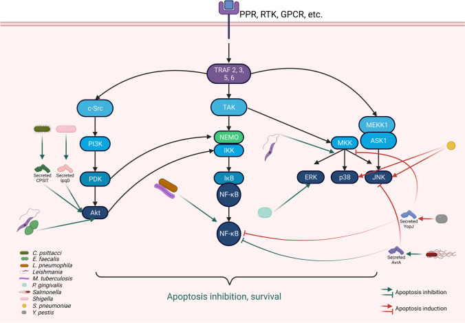
Apoptosis is a finely programmed process of cell death in which cells silently dismantle and actively participate in several operations such as immune response, differentiation, and cell growth. It can be initiated by three main pathways: the extrinsic, the perforin granzyme, and the intrinsic that culminate in the activation of several proteins in charge of tearing down the cell. On the other hand, apoptosis represents an ordeal for pathogens that live inside cells and maintain a strong dependency with them; thus, they have evolved multiple strategies to manipulate host cell apoptosis on their behalf. It has been widely documented that diverse intracellular bacteria, fungi, and parasites can interfere with most steps of the host cell apoptotic machinery to inhibit or induce apoptosis. Indeed, the inhibition of apoptosis is considered a virulence property shared by many intracellular pathogens to ensure productive replication. Some pathogens intervene at an early stage by interfering with the sensing of extracellular signals or transduction pathways. Others sense cellular stress or target the apoptosis regulator proteins of the Bcl-2 family or caspases. In many cases, the exact molecular mechanisms leading to the interference with the host cell apoptotic cascade are still unknown. However, intense research has been conducted to elucidate the strategies employed by intracellular pathogens to modulate host cell death. In this review, we summarize the main routes of activation of apoptosis and present several processes used by different bacteria, fungi, and parasites to modulate the apoptosis of their host cells.
Keywords: Apoptosis; Bacteria; Fungi; Intracellular pathogens; Parasites; Signaling pathways.
© 2023. The Author(s).
Conflict of interest statement
The authors declare no competing interests.
Figures


Similar articles
-
Modulation of host cell apoptotic pathways by intracellular pathogens.Curr Opin Microbiol. 2017 Feb;35:88-99. doi: 10.1016/j.mib.2017.03.001. Epub 2017 Mar 19. Curr Opin Microbiol. 2017. PMID: 28319728 Review.
-
Inhibition of apoptosis by intracellular protozoan parasites.Int J Parasitol. 2001 Sep;31(11):1166-76. doi: 10.1016/s0020-7519(01)00271-5. Int J Parasitol. 2001. PMID: 11563357 Review.
-
Inflammasomes, Autophagy, and Cell Death: The Trinity of Innate Host Defense against Intracellular Bacteria.Mediators Inflamm. 2019 Jan 8;2019:2471215. doi: 10.1155/2019/2471215. eCollection 2019. Mediators Inflamm. 2019. PMID: 30728749 Free PMC article. Review.
-
The regulation of apoptosis by microbial pathogens.Int Rev Cytol. 1999;187:203-59. doi: 10.1016/s0074-7696(08)62419-5. Int Rev Cytol. 1999. PMID: 10212981 Review.
-
The complexity of apoptotic cell death in mollusks: An update.Fish Shellfish Immunol. 2015 Sep;46(1):79-87. doi: 10.1016/j.fsi.2015.03.038. Epub 2015 Apr 9. Fish Shellfish Immunol. 2015. PMID: 25862972 Review.
Cited by
-
Preparation of Iron oxide/palladium nanoparticles modified with carbon quantum dots (Pd@CQD@Fe3O4): insight to anti-cancer effect and ROS generation.Sci Rep. 2025 May 19;15(1):17317. doi: 10.1038/s41598-025-02386-0. Sci Rep. 2025. PMID: 40389602 Free PMC article.
-
Beyond survival to domination: Brucella's multilayered strategies for evading host immune responses.Front Microbiol. 2025 Jun 18;16:1608617. doi: 10.3389/fmicb.2025.1608617. eCollection 2025. Front Microbiol. 2025. PMID: 40606156 Free PMC article. Review.
-
Identification of the Francisella novicida FTN_0096 as a factor involved in intracellular replication and host response.PLoS One. 2025 Aug 1;20(8):e0329626. doi: 10.1371/journal.pone.0329626. eCollection 2025. PLoS One. 2025. PMID: 40748985 Free PMC article.
-
Engineering of the Caspase-3 Gene in Recombinant CHO Cells Caused More Apoptosis Resistance and enhanced Recombinant Protein Production Than the BAX Gene.Iran Biomed J. 2025 May 1;29(3):149-158. doi: 10.61186/ibj.4934. Iran Biomed J. 2025. PMID: 40588867 Free PMC article.
-
Host factor RBMX2 promotes epithelial cell apoptosis by downregulating APAF-1's Retention Intron after Mycobacterium bovis infection.Front Immunol. 2024 Sep 6;15:1431207. doi: 10.3389/fimmu.2024.1431207. eCollection 2024. Front Immunol. 2024. PMID: 39308873 Free PMC article.
References
Publication types
MeSH terms
Substances
LinkOut - more resources
Full Text Sources
