

Exploration of inorganic nanoparticles for revolutionary drug delivery applications: a critical review
- PMID: 38112849
- PMCID: PMC10730791
- DOI: 10.1186/s11671-023-03943-0
Exploration of inorganic nanoparticles for revolutionary drug delivery applications: a critical review
Abstract
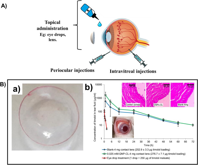
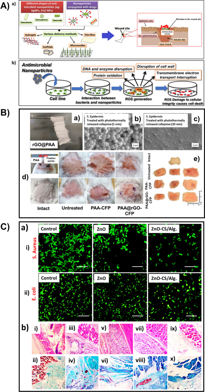
The nanosystems for delivering drugs which have evolved with time, are being designed for greater drug efficiency and lesser side-effects, and are also complemented by the advancement of numerous innovative materials. In comparison to the organic nanoparticles, the inorganic nanoparticles are stable, have a wide range of physicochemical, mechanical, magnetic, and optical characteristics, and also have the capability to get modified using some ligands to enrich their attraction towards the molecules at the target site, which makes them appealing for bio-imaging and drug delivery applications. One of the strong benefits of using the inorganic nanoparticles-drug conjugate is the possibility of delivering the drugs to the affected cells locally, thus reducing the side-effects like cytotoxicity, and facilitating a higher efficacy of the therapeutic drug. This review features the direct and indirect effects of such inorganic nanoparticles like gold, silver, graphene-based, hydroxyapatite, iron oxide, ZnO, and CeO2 nanoparticles in developing effective drug carrier systems. This article has remarked the peculiarities of these nanoparticle-based systems in pulmonary, ocular, wound healing, and antibacterial drug deliveries as well as in delivering drugs across Blood-Brain-Barrier (BBB) and acting as agents for cancer theranostics. Additionally, the article sheds light on the plausible modifications that can be carried out on the inorganic nanoparticles, from a researcher's perspective, which could open a new pathway.
Keywords: Blood–brain-barrier; Drug delivery; Inorganic nanoparticles; Theranostics; Wound healing.
© 2023. The Author(s).
Conflict of interest statement
The authors declare no competing interests.
Figures
Similar articles
-
Engineered inorganic nanoparticles for drug delivery applications.Curr Drug Metab. 2013 Jun;14(5):518-30. doi: 10.2174/13892002113149990008. Curr Drug Metab. 2013. PMID: 23116108 Review.
-
Inorganic-organic Synergy in Nano-hybrids Makes a New Class of Drug with Targeted Delivery: Glutamate Functionalization of Iron Nanoparticles for Potential Bone Marrow Delivery and X-ray Dynamic Therapy.Curr Drug Deliv. 2022;19(10):991-1000. doi: 10.2174/1567201819666220328142620. Curr Drug Deliv. 2022. PMID: 35346006 Review.
-
Recent Progress in Delivery of Therapeutic and Imaging Agents Utilizing Organic-Inorganic Hybrid Nanoparticles.Curr Drug Deliv. 2018;15(4):485-496. doi: 10.2174/1567201814666171120114034. Curr Drug Deliv. 2018. PMID: 29165073 Review.
-
Optically active organic and inorganic nanomaterials for biological imaging applications: A review.Micron. 2023 Sep;172:103486. doi: 10.1016/j.micron.2023.103486. Epub 2023 May 24. Micron. 2023. PMID: 37262930 Review.
-
Inorganic Nanoparticles for Cancer Therapy: A Transition from Lab to Clinic.Curr Med Chem. 2018;25(34):4269-4303. doi: 10.2174/0929867325666171229141156. Curr Med Chem. 2018. PMID: 29284391 Review.
Cited by
-
Nanotechnology in Parkinson's Disease: overcoming drug delivery challenges and enhancing therapeutic outcomes.Drug Deliv Transl Res. 2025 Jan 29. doi: 10.1007/s13346-025-01799-8. Online ahead of print. Drug Deliv Transl Res. 2025. PMID: 39878857
-
Unlocking the potential of remdesivir: innovative approaches to drug delivery.Drug Deliv Transl Res. 2025 Apr 17. doi: 10.1007/s13346-025-01843-7. Online ahead of print. Drug Deliv Transl Res. 2025. PMID: 40244526 Review.
-
Nanostructured Formulations for a Local Treatment of Cancer: A Mini Review About Challenges and Possibilities.Pharmaceutics. 2025 Feb 6;17(2):205. doi: 10.3390/pharmaceutics17020205. Pharmaceutics. 2025. PMID: 40006574 Free PMC article. Review.
-
Rational Design of Safer Inorganic Nanoparticles via Mechanistic Modeling-Informed Machine Learning.ACS Nano. 2025 Jun 17;19(23):21538-21555. doi: 10.1021/acsnano.5c03590. Epub 2025 Jun 3. ACS Nano. 2025. PMID: 40460056 Free PMC article.
-
Postbiotics: Modulation of the Gut Microbiota and Potential for Association with Nanotechnology.Probiotics Antimicrob Proteins. 2025 Jul 18. doi: 10.1007/s12602-025-10675-3. Online ahead of print. Probiotics Antimicrob Proteins. 2025. PMID: 40679568 Review.
References
-
- Shah S, Patel AA, Prajapati BG, Alexander A, Pandya V, Trivedi N, Pandey P, Patel SG, Patel RJ. Multifaceted nanolipidic carriers: a modish stratagem accentuating nose-to-brain drug delivery. J Nanopart Res. 2023;25:150. doi: 10.1007/s11051-023-05804-4. - DOI
-
- Megha M, Joy A, Unnikrishnan G, Jayan M, Haris M, Thomas J, Kolanthai E, Muthuswamy S. Structural and biological evaluation of novel vanadium/Yttrium co-doped hydroxyapatite for bone tissue engineering applications. J Alloys Compd. 2023;967:171697. doi: 10.1016/j.jallcom.2023.171697. - DOI
-
- Megha M, Joy A, Unnikrishnan G, Haris M, Thomas J, Deepti A, Chakrapani PSB, Kolanthai E, Muthuswamy S. Structural and biological properties of novel Vanadium and Strontium co-doped HAp for tissue engineering applications. Ceram Int. 2023;49:30156–30169. doi: 10.1016/j.ceramint.2023.06.272. - DOI
Publication types
LinkOut - more resources
Full Text Sources