. 2024 Mar 4;12(3):350-362.
doi: 10.1158/2326-6066.CIR-23-0496.

LILRB3 Supports Immunosuppressive Activity of Myeloid Cells and Tumor Development

Affiliations

LILRB3 Supports Immunosuppressive Activity of Myeloid Cells and Tumor Development

Ryan Huang et al. Cancer Immunol Res. .

Abstract

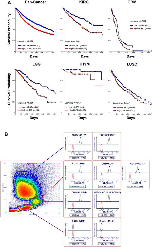
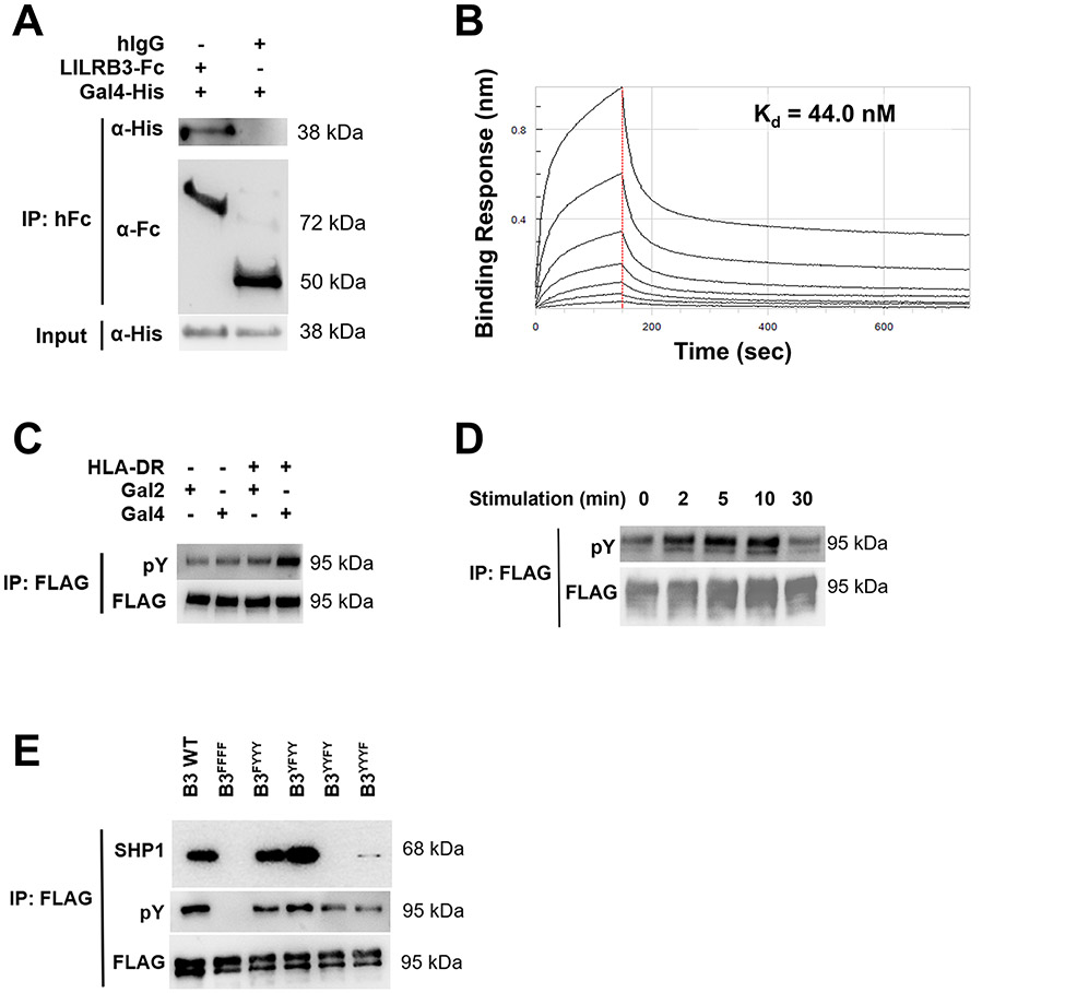
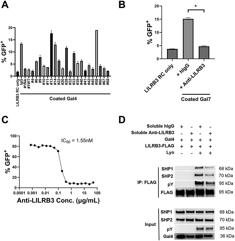
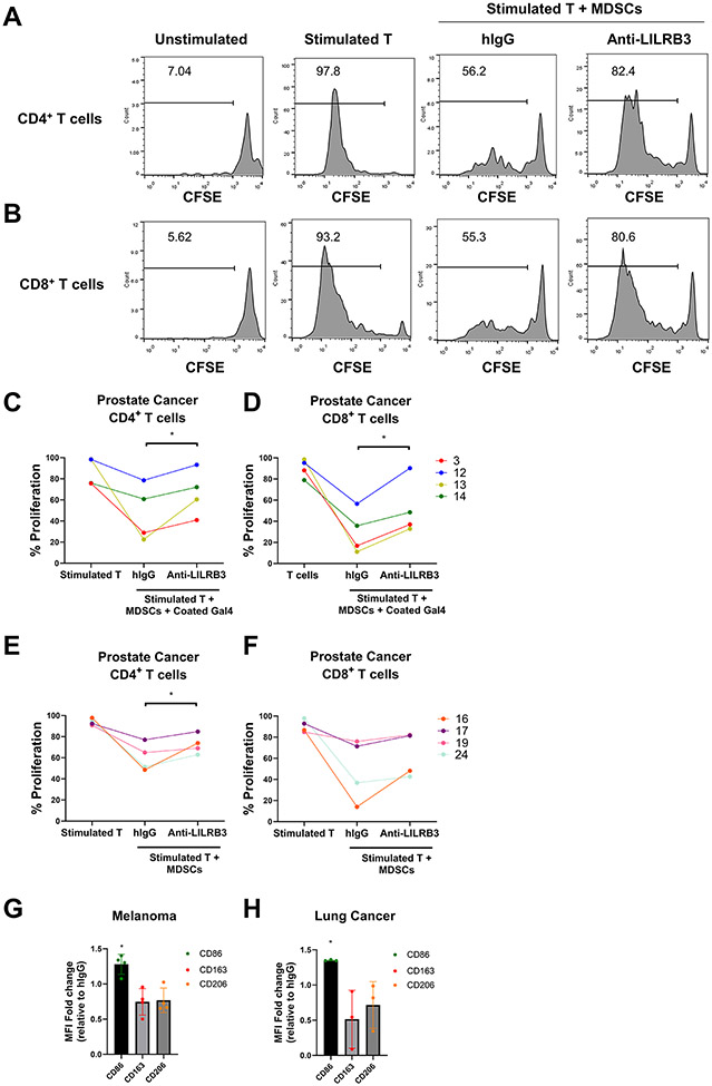
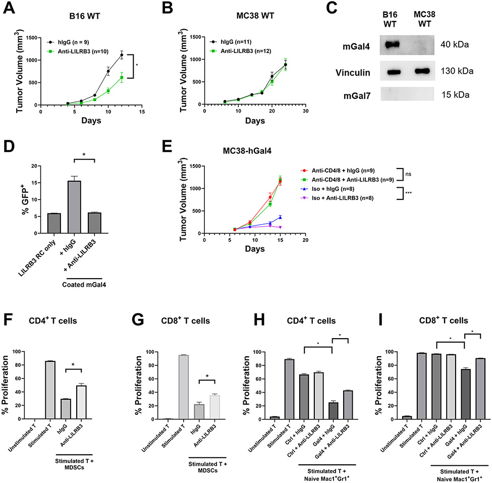
The existing T cell-centered immune checkpoint blockade therapies have been successful in treating some but not all patients with cancer. Immunosuppressive myeloid cells, including myeloid-derived suppressor cells (MDSC), that inhibit antitumor immunity and support multiple steps of tumor development are recognized as one of the major obstacles in cancer treatment. Leukocyte Ig-like receptor subfamily B3 (LILRB3), an immune inhibitory receptor containing tyrosine-based inhibitory motifs (ITIM), is expressed solely on myeloid cells. However, it is unknown whether LILRB3 is a critical checkpoint receptor in regulating the activity of immunosuppressive myeloid cells, and whether LILRB3 signaling can be blocked to activate the immune system to treat solid tumors. Here, we report that galectin-4 and galectin-7 induce activation of LILRB3 and that LILRB3 is functionally expressed on immunosuppressive myeloid cells. In some samples from patients with solid cancers, blockade of LILRB3 signaling by an antagonistic antibody inhibited the activity of immunosuppressive myeloid cells. Anti-LILRB3 also impeded tumor development in myeloid-specific LILRB3 transgenic mice through a T cell-dependent manner. LILRB3 blockade may prove to be a novel approach for immunotherapy of solid cancers.


Conflict of interest statement

Conflict of interest statement: Authors R.H., X.L., J.K., M.D., H.C., G.W., X.G., J.X., Y.H., S.J., N.Z., Z.A., and C.C.Z. are listed as inventors in LILRB relevant patent applications that were exclusively licensed to Immune-Onc Therapeutic, Inc. by the Board of Regents of the University of Texas System. Authors Z.A., N.Z., and C.C.Z. hold equity in and have Sponsored Research Agreements with Immune-Onc Therapeutics, Inc. University of Texas has a financial interest in Immune-Onc in the form of equity and licensing.

Figures

Fig. 1∣
Fig. 1∣. LILRB3 expression is associated with reduced survival in patients diagnosed with solid tumors.
(a) Kaplan-Meier survival analysis of correlations between LILRB3 and overall survival of patients from the TCGA database. Patients were stratified with low levels of LILRB3 (blue) or high levels of LILRB3 (red) from TCGA subsets of pan-cancer, renal clear cell carcinoma (KIRC), glioblastoma (GBM), low-grade glioma (LGG), thymoma (THYM), and lung squamous cell carcinoma (LUSC). Statistical significance for overall survival was calculated by Mann-Whitney’s log-rank test. (b) Expression of LILRB3 on the cell surface of different subsets of lymphocytes, monocytes, and granulocytes in a representative human patient with melanoma by flow cytometry.
Fig. 2∣
Fig. 2∣. Galectin-4 activates LILRB3 in a site-specific manner.
(a) Percentages of indicated LILRB reporter cells activated by K562-Vec or K562-HLA-G cells. (b-c) Percentages of LILRB3 (b) or LILRB4 (c) reporter cells activated on recombinant coated ApoE or coated anti-LILRB3 (20 μg/mL). (d-h) Percentages of LILRB1 (d), LILRB2 (e), LILRB3 (f), LILRB4 (g), or LILRB5 (h) reporter cells activated on coated galectins (20 μg/mL). (i) Summary of LILRB reporter cells activated on coated galectins. (j) Percentages of LILRB3 reporter cells activated on coated or soluble galectin-4 or galectin-7 (20 μg/mL). (k) Percentages of LILRB31-443 or LILRB31-419 reporter cells activated on coated galectin-4 or anti-LILRB3 (20 μg/mL). Three technical replicates were performed for each condition and at least two independent experiments were performed for each condition. Error bars represent SEM and * indicates two-tailed student’s t-test p < 0.05.
Fig. 3∣
Fig. 3∣. Galectin-4 binds LILRB3 and induces downstream signaling in an ITIM-dependent manner.
(a) Co-immunoprecipitation (Co-IP) demonstrates that recombinant Fc-tagged extracellular domain of LILRB3 (LILRB3-ECD) but not human IgG (hIgG) attached to Protein A Dynabeads interacts with recombinant human galectin-4 (hGal4). (b) Binding kinetics of His-tagged human galectin-4 to immobilized LILRB3-ECD were measured using biolayer interferometry and determined using a 1:1 binding curve. (c) Representative Co-IP of LILRB3-specific phosphotyrosine (pY) Western blot of THP1-LILRB3 lysates after 10 minutes on coated HLA-DR (20 μg/mL) and coated human galectin-2 (20 μg/mL) or human galectin-4 (20 μg/mL). (d) Representative Co-IP of LILRB3-specific pY Western blot of THP1-LILRB3 lysates after indicated time points with soluble LPS (200 ng/mL) and coated and soluble human galectin-4 (20 μg/mL). (e) Representative Co-IP of LILRB3-specific pY and SHP1 Western blot of 293T cells transfected with SHP1/SHP2/hGal4/Lyn and LILRB3-WT or LILRB3 ITIM-mutated plasmids after 48 hours. Three technical replicates were performed for each condition and at least two independent experiments were performed for each condition.
Fig. 4∣
Fig. 4∣. Anti-LILRB3 antibody blocks Galectin-4 induced SHP1/SHP2 signaling through LILRB3.
(a) Percentages of LILRB3 reporter cells activated on coated human galectin-4 (20 μg/mL) with indicated soluble anti-LILRB3 antibodies (20 μg/mL). (b) Percentages of LILRB3 reporter cells activated on coated human galectin-7 (20 μg/mL) with soluble hIgG control or anti-LILRB3-1 (20 μg/mL). (c) Percentages of LILRB3 reporter cells activated on coated human Galectin-4 (20 μg/mL) with indicated concentrations of soluble anti-LILRB3-1. (d) Representative Co-IP of full-length LILRB3-specific pY/SHP1/SHP2 Western blot of 293T cells transfected with SHP1/SHP2/hGal4/LILRB3-WT/Lyn plasmids with soluble hIgG control or anti-LILRB3-1 antibody. Three technical replicates were performed for each condition and at least two independent experiments were performed for each condition. Error bars represent SEM and * indicates two-tailed student’s t-test p < 0.05.
Fig. 5∣
Fig. 5∣. Anti-LILRB3 antibody inhibits human MDSC activity.
(a-b) Representative histogram of 31 patients with cancer (total of 76 patients with cancer) shows that anti-LILRB3 attenuates MDSC suppressive functions on CD4+ (a) and CD8+ (b) T-cell proliferation in some patients with cancer as determined by flow cytometry. (c-d) Quantification of 4 individual patient samples from proliferating human CD4+ (c) and CD8+ (d) T cells cocultured with autologous MDSCs selected for patients with >10% proliferation with anti-LILRB3. Wells were coated with human galectin-4 (20 μg/mL) and cocultured cells were treated with hIgG control or anti-LILRB3 (20 μg/mL). (e-f) Quantification of 4 individual patient samples from proliferating human CD4+ (e) and CD8+ (f) T cells cultured with autologous MDSCs selected for patients with >10% proliferation with anti-LILRB3. Wells were not coated with human galectin-4 and cocultured cells were treated with hIgG control or anti-LILRB3 (20 μg/mL). (g-h) Median fluorescence intensity (MFI) change in CD86, CD163, and CD206 for isolated MDSCs treated with hIgG control or anti-LILRB3 (20 μg/mL) in patient samples with melanoma (g) or lung cancer (h). One to three replicates were performed for each condition and one experiment was performed for each condition. Error bars represent SEM and * indicates two-tailed student’s t-test p < 0.05.
Fig. 6∣
Fig. 6∣. Anti-LILRB3 antibody ameliorates cancer development in vivo.
(a-b) Tumor progression of LILRB3 transgenic mice challenged with 5X10⁵ B16 melanoma cells (a) or MC38 colon cancer cells (b) and treated with either hIgG control or anti-LILRB3. (c) Representative Western blot of B16 and MC38 whole cell lysates for mouse galectin-4, vinculin loading control, and mouse galectin-7. (d) Percentages of LILRB3 reporter cells activated on coated mouse galectin-4 (20 μg/mL) with hIgG control or anti-LILRB3 (20 μg/mL). (e) Tumor progression of LILRB3 transgenic mice challenged with 5X10⁵ MC38-hGal4 and treated with hIgG control or anti-LILRB3, and a combination of anti-CD4 and anti-CD8 or isotype control. (f-g) Quantification of proliferating mouse CD4+ (f) or CD8+ (g) T cells cocultured with MDSCs from B16-challenged LILRB3 transgenic mice. Cocultured cells were treated with hIgG control or anti-LILRB3 (20 μg/mL). (h-i) Quantification of proliferating mouse CD4+ (h) or CD8+ (i) T cells cocultured with naïve Mac1+Gr1+ cells from unchallenged LILRB3 transgenic mice on coated human galectin-4 or BSA (10 μg/mL) and treated with hIgG control or anti-LILRB3 (20 μg/mL). Three technical replicates were performed for each condition and at least two independent experiments were performed for each condition. Error bars represent SEM and * indicates two-tailed student’s t-test p < 0.05. *** indicates two-tailed student’s t-test p < 0.0005.

References

    1. Barry ST, Gabrilovich DI, Sansom OJ, Campbell AD, Morton JP. Therapeutic targeting of tumour myeloid cells. Nat Rev Cancer 2023;23(4):216–37 doi 10.1038/s41568-022-00546-2. - DOI - PubMed
    1. Salemme V, Centonze G, Cavallo F, Defilippi P, Conti L. The Crosstalk Between Tumor Cells and the Immune Microenvironment in Breast Cancer: Implications for Immunotherapy. Front Oncol 2021;11:610303 doi 10.3389/fonc.2021.610303. - DOI - PMC - PubMed
    1. Samaridis J, Colonna M. Cloning of novel immunoglobulin superfamily receptors expressed on human myeloid and lymphoid cells: structural evidence for new stimulatory and inhibitory pathways. Eur J Immunol 1997;27(3):660–5 doi 10.1002/eji.1830270313. - DOI - PubMed
    1. Kang X, Kim J, Deng M, John S, Chen H, Wu G, et al. Inhibitory leukocyte immunoglobulin-like receptors: Immune checkpoint proteins and tumor sustaining factors. Cell Cycle 2016;15(1):25–40 doi 10.1080/15384101.2015.1121324. - DOI - PMC - PubMed
    1. Hirayasu K, Arase H. Functional and genetic diversity of leukocyte immunoglobulin-like receptor and implication for disease associations. Journal of human genetics 2015;60(11):703–8 doi 10.1038/jhg.2015.64. - DOI - PubMed

Publication types