Enlarged Perivascular Spaces in Infancy and Autism Diagnosis, Cerebrospinal Fluid Volume, and Later Sleep Problems
- PMID: 38113043
- PMCID: PMC10731509
- DOI: 10.1001/jamanetworkopen.2023.48341
Enlarged Perivascular Spaces in Infancy and Autism Diagnosis, Cerebrospinal Fluid Volume, and Later Sleep Problems
Abstract
Importance: Perivascular spaces (PVS) and cerebrospinal fluid (CSF) are essential components of the glymphatic system, regulating brain homeostasis and clearing neural waste throughout the lifespan. Enlarged PVS have been implicated in neurological disorders and sleep problems in adults, and excessive CSF volume has been reported in infants who develop autism. Enlarged PVS have not been sufficiently studied longitudinally in infancy or in relation to autism outcomes or CSF volume.
Objective: To examine whether enlarged PVS are more prevalent in infants who develop autism compared with controls and whether they are associated with trajectories of extra-axial CSF volume (EA-CSF) and sleep problems in later childhood.
Design, setting, and participants: This prospective, longitudinal cohort study used data from the Infant Brain Imaging Study. Magnetic resonance images were acquired at ages 6, 12, and 24 months (2007-2017), with sleep questionnaires performed between ages 7 and 12 years (starting in 2018). Data were collected at 4 sites in North Carolina, Missouri, Pennsylvania, and Washington. Data were analyzed from March 2021 through August 2022.
Exposure: PVS (ie, fluid-filled channels that surround blood vessels in the brain) that are enlarged (ie, visible on magnetic resonance imaging).
Main outcomes and measures: Outcomes of interest were enlarged PVS and EA-CSF volume from 6 to 24 months, autism diagnosis at 24 months, sleep problems between ages 7 and 12 years.
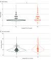
Results: A total of 311 infants (197 [63.3%] male) were included: 47 infants at high familial likelihood for autism (ie, having an older sibling with autism) who were diagnosed with autism at age 24 months, 180 high likelihood infants not diagnosed with autism, and 84 low likelihood control infants not diagnosed with autism. Sleep measures at school-age were available for 109 participants. Of infants who developed autism, 21 (44.7%) had enlarged PVS at 24 months compared with 48 infants (26.7%) in the high likelihood but no autism diagnosis group (P = .02) and 22 infants in the control group (26.2%) (P = .03). Across all groups, enlarged PVS at 24 months was associated with greater EA-CSF volume from ages 6 to 24 months (β = 4.64; 95% CI, 0.58-8.72; P = .002) and more frequent night wakings at school-age (F = 7.76; η2 = 0.08; P = .006).
Conclusions and relevance: These findings suggest that enlarged PVS emerged between ages 12 and 24 months in infants who developed autism. These results add to a growing body of evidence that, along with excessive CSF volume and sleep dysfunction, the glymphatic system could be dysregulated in infants who develop autism.
Conflict of interest statement
Figures


Comment in
-
Beyond the AJR: Updates on the Glymphatic System, Perivascular Spaces, and Autism During Infancy.AJR Am J Roentgenol. 2024 Dec;223(6):e2431220. doi: 10.2214/AJR.24.31220. Epub 2024 Apr 17. AJR Am J Roentgenol. 2024. PMID: 38630087 No abstract available.
References
Publication types
MeSH terms
Grants and funding
LinkOut - more resources
Full Text Sources
Research Materials

