. 2023 Dec;16(12):e010351.
doi: 10.1161/CIRCHEARTFAILURE.122.010351. Epub 2023 Dec 19.

Nonsense Variant PRDM16-Q187X Causes Impaired Myocardial Development and TGF-β Signaling Resulting in Noncompaction Cardiomyopathy in Humans and Mice

Affiliations

Nonsense Variant PRDM16-Q187X Causes Impaired Myocardial Development and TGF-β Signaling Resulting in Noncompaction Cardiomyopathy in Humans and Mice

Bo Sun et al. Circ Heart Fail. 2023 Dec.

Abstract

Background: PRDM16 plays a role in myocardial development through TGF-β (transforming growth factor-beta) signaling. Recent evidence suggests that loss of PRDM16 expression is associated with cardiomyopathy development in mice, although its role in human cardiomyopathy development is unclear. This study aims to determine the impact of PRDM16 loss-of-function variants on cardiomyopathy in humans.

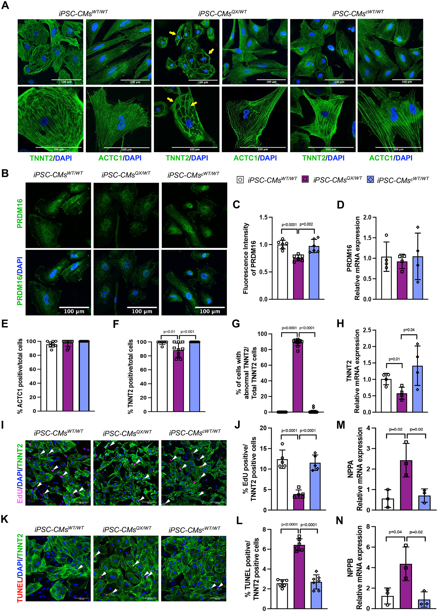
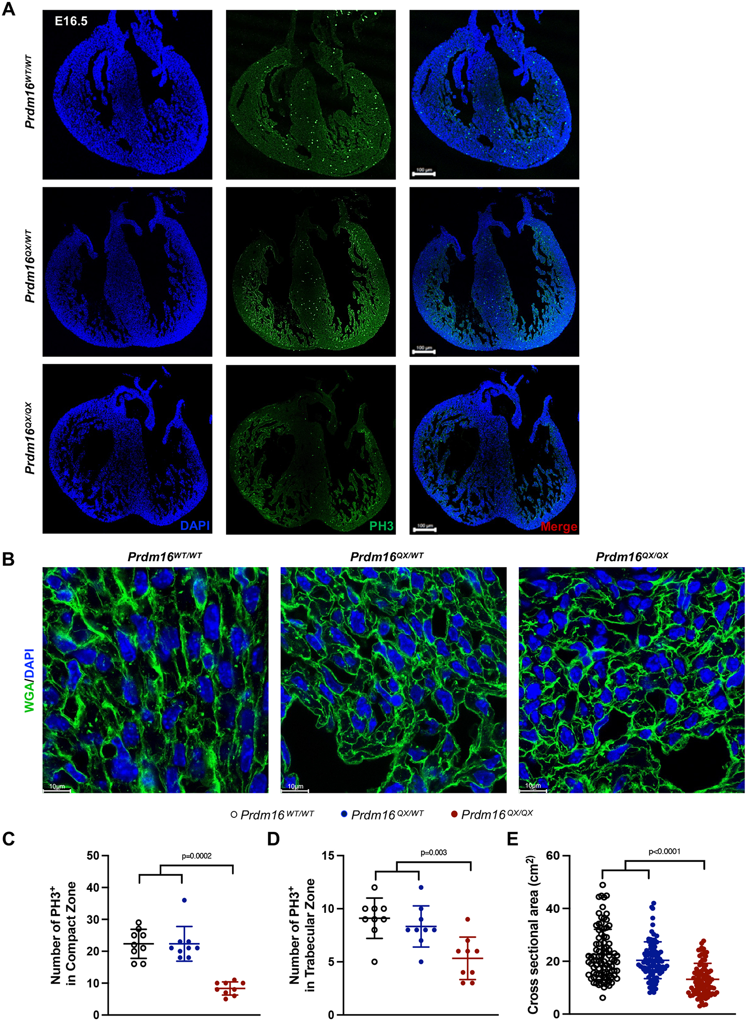
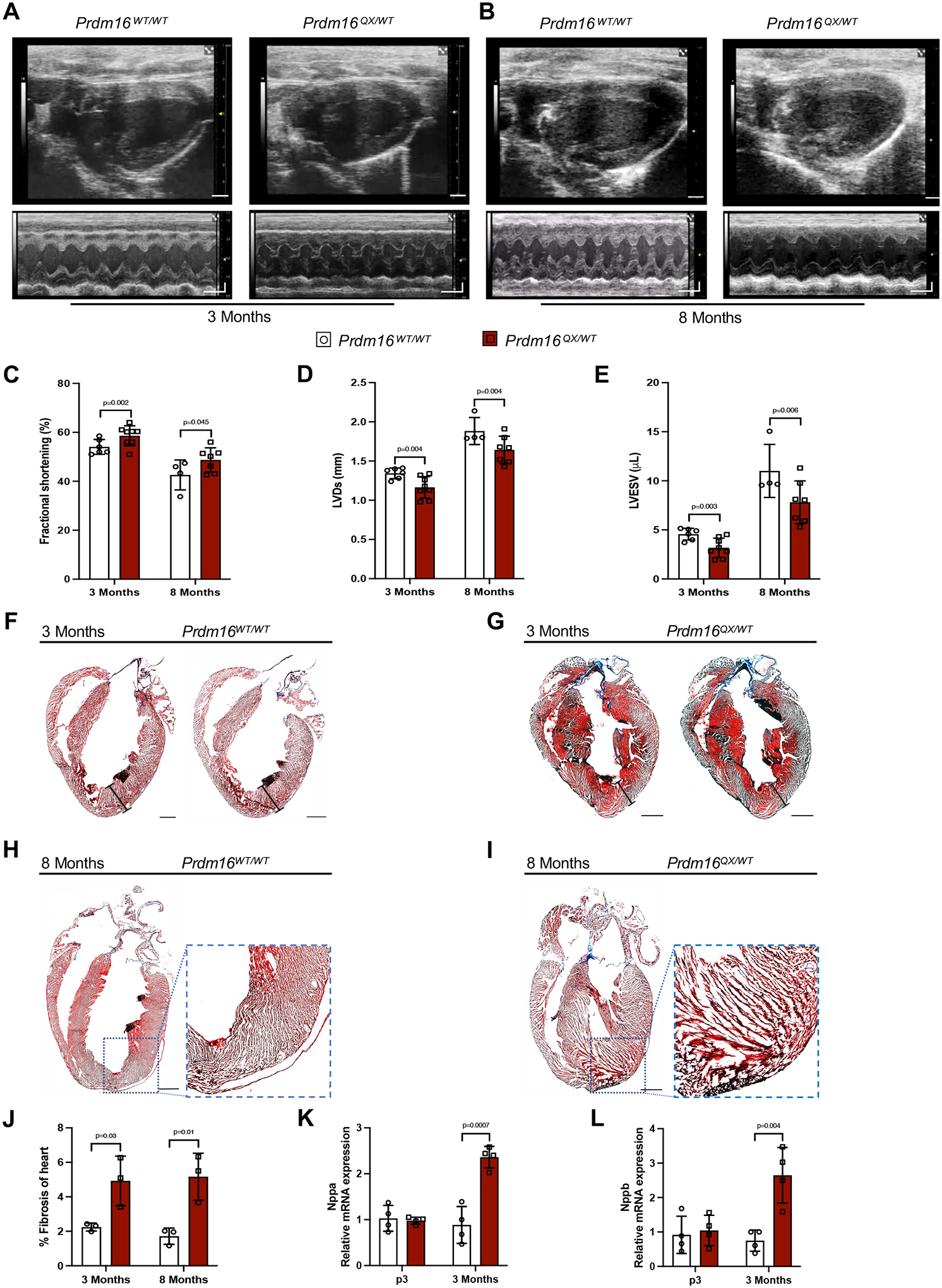
Methods: Individuals with PRDM16 variants were identified and consented. Induced pluripotent stem cell-derived cardiomyocytes were generated from a proband hosting a Q187X nonsense variant as an in vitro model and underwent proliferative and transcriptional analyses. CRISPR (clustered regularly interspaced short palindromic repeats)-mediated knock-in mouse model hosting the Prdm16Q187X allele was generated and subjected to ECG, histological, and transcriptional analysis.

Results: We report 2 probands with loss-of-function PRDM16 variants and pediatric left ventricular noncompaction cardiomyopathy. One proband hosts a PRDM16-Q187X variant with left ventricular noncompaction cardiomyopathy and demonstrated infant-onset heart failure, which was selected for further study. Induced pluripotent stem cell-derived cardiomyocytes prepared from the PRDM16-Q187X proband demonstrated a statistically significant impairment in myocyte proliferation and increased apoptosis associated with transcriptional dysregulation of genes implicated in cardiac maturation, including TGF-β-associated transcripts. Homozygous Prdm16Q187X/Q187X mice demonstrated an underdeveloped compact myocardium and were embryonically lethal. Heterozygous Prdm16Q187X/WT mice demonstrated significantly smaller ventricular dimensions, heightened fibrosis, and age-dependent loss of TGF-β expression. Mechanistic studies were undertaken in H9c2 cardiomyoblasts to show that PRDM16 binds TGFB3 promoter and represses its transcription.

Conclusions: Novel loss-of-function PRDM16 variant impairs myocardial development resulting in noncompaction cardiomyopathy in humans and mice associated with altered TGF-β signaling.

Keywords: alleles; apoptosis; cardiomyopathies; induced pluripotent stem cells; mice; transcription factors.


Conflict of interest statement

Disclosures None.

Figures

Figure 1:
Figure 1:
Loss-of-function genetics variants in PRDM16 are associated with left ventricular noncompaction cardiomyopathy (LVNC). (a) Schematic pedigree of Family A with LVNC. Proband 1 (III.1, arrow) hosts a heterozygous nonsense mutation (PRDM16-Q187X) denoted with a + and has LVNC with pediatric onset heart failure. DCM, dilated cardiomyopathy. SVT, supraventricular tachycardia. (b) Echocardiogram of Proband 1 demonstrates trabeculations of the left ventricular apex consistent with LVNC (yellow arrowheads) present at 3 days of life that progressed to DCM by 3 months of age. (c) Sequence chromatogram of genotype wild-type III.2 and genotype PRDM16-Q187X III.1 kindred with the mutated nucleotide noted with the arrow. (d) Western blot of empty vector (EV), human PRDM16 wild-type (WT), and PRDM16-Q187X protein (QX) overexpressed in HEK293T cells. (e) Schematic pedigree Family B with LVNC. Proband 2 (II.1, arrow) hosts a heterozygous splice variant (c.676+2T) in PRDM16. (f) Echocardiogram of Proband 2 (II.1) showing prominent apical trabeculations (yellow arrowheads) consistent with LVNC.
Figure 2:
Figure 2:
PRDM16-Q187X impairs proliferation and increases apoptosis in iPSC-derived cardiomyocytes (iPSC-CMs). (a) Representative images of iPSC-CM differentiation markers Troponin T (TNNT2) and cardiac muscle alpha actin (ACTC1) in iPSC-CMs from non-isogenic control (iPSC-CMsWT/WT), Proband 1 (iPSC-CMsQX/WT) and CRISPR-corrected isogenic control (iPSC-CMscWT/WT). Yellow arrowheads demonstrated an abnormal organization of TNNT2 with loss of parallel myofilament alignment. (b) Immunostaining of nuclei (blue) and PRDM16 (green) in iPSC-CMs. (c) Fluorescence intensity of PRDM16 in iPSC-CMs (n=3). (d) PRDM16 mRNA expression in Proband 1 (iPSC-CMsQX/WT) compared with controls (n=4). (e) Quantification of ACTC1 expression in iPSC-CMs (n=3). (f) Quantification of TNNT2 expression in iPSC-CMs (n=3). (g) Quantification of cells with abnormal TNNT2 pattern over the total number of cells (n=3). (h) Relative mRNA expression (normalized to RPL32) of TNNT2 from iPSC-CMs (n=4). (i) Immunostaining of nuclei (blue), TNNT2 (green) and EdU (pink) in iPSC-CMs at 4 weeks. White arrowheads represent EdU positive cells. (j) Percentage of EdU positive cardiomyocytes in Proband 1 and controls (n=3). (k) Immunostaining of nuclei (blue), TNNT2 (green) and TUNEL (red) in iPSC-CMs at 4 weeks. White arrowheads represent TUNEL positive cells. (l) Percentage of TUNEL iPSC-CMs (n=3). (m, n) mRNA expression of NPPA (encoding natriuretic peptide A) and NPPB (encoding natriuretic peptide B) in iPSC-CMs (n=3). The bar graphs show the mean and error bars represent ± SD. Scale bar: 100 μm. P values generated using unpaired t-test.
Figure 3:
Figure 3:
Prdm16QX/QX homozygous mice display left ventricular compaction defect: (a-c) Sequence chromatograms confirming the genotype of control (Prdm16WT/WT), heterozygote (Prdm16QX/WT) and homozygous mutant (Prdm16QX/QX) knock-in mice, respectively. NruI, Bsp68I restriction enzyme. (d) Prdm16WT/WT, Prdm16WT/QX and Prdm16QX/QX E16.5 hearts were stained with endomucin (Endo) to label endocardial cell and Ki67 to identify proliferative cells. (e-f) Quantification of the thickness of noncompacted zone and the noncompacted/compacted ratio (at least three hearts per genotype, 6 fields were measured per section). (g-h) Cardiomyocyte proliferation rate using average of Ki67+ cells in six field of view (at least three hearts per genotype). (i) Quantification of left ventricle thickness (at least three hearts per genotype, 6 different spots were measured per section). The bar graphs show the mean and error bars represent ± SD. Scale bars:100 μm. P-values generated using one-way ANOVA (not shown) followed by Tukey post hoc test.
Figure 4:
Figure 4:
Prdm16QX/QX homozygous mice display defects in cardiomyocyte development. (a) Prdm16WT/WT, Prdm16WT/QX, Prdm16QX/QX E16.5 hearts were stained with anti-histone H3 (PH3) to quantify proliferative cells. Scale bars:100 μm. (b) Representative WGA (Wheat germ agglutinin) staining images for visualizing cell size. Scale bars: 10um. (c, d) Quantification of PH3 positive (pH3+) cells in both compact and trabecular areas of the heart show a significant decrease in cardiomyocyte proliferation rate in Prdm16QX/QX when compared to Prdm16WT/WT, Prdm16QX/WT (calculated from at least 3 sections). (e) Quantification of cross-sectional area shows a significant decrease in cell size in Prdm16QX/QX compared to Prdm16WT/WT, Prdm16QX/WT (n=3 sections, 90 cardiomyocytes/section/group). The bar graphs show the mean and error bars represent ± SD. P-values generated using one-way ANOVA (not shown) followed by Tukey post hoc test.
Figure 5:
Figure 5:
Prdm16QX/WT mutant mice develop pathological cardiac remodeling. (a, b) Long-axis B-mode and M-mode echocardiographic images at 3 and 8 months, respectively. Scale bar:100 ms. (c) Ejection fraction (n=4, 6, 7 and 8 mice per group, each dot corresponds to a mouse); (d) Left ventricular diameter in systole (LVDs) (n=4, 6, 7 and 8 mice per group, each dot corresponds to a mouse); (e) Left ventricular end systolic volume (LVESV) in Prdm16WT/WT and Prdm16QX/WT mice at 3 and 8 months of age (n=4, 6, 7 and 8 mice per group, each dot corresponds to a mouse); (f-i) Histological analysis of heart sections stained with hematoxylin and eosin or trichrome from 3 and 8 months-old mice showing small left ventricular (LV) size and internal dimensions. Scale bars: 1000 μm. (j) Quantification of the amount of fibrosis in the heart at 3 and 8 months, respectively (n=3 hearts). (k, l) Quantification of mRNA expression of Nppa (encoding natriuretic peptide A) and Nppb (encoding natriuretic peptide B) in hearts of 3 days (p3) and 3 months-old mice (n=4 hearts). The bar graphs show the mean and error bars represent ± SD. Comparisons are by unpaired t-tests.
Figure 6:
Figure 6:
PRDM16-Q187X is associated with modulation of developmentally-dependent Tgfβ signaling. (a) Heatmap of differentially expressed transcripts from RNA-seq analysis of mutant iPSC-cardiac myocytes (iPSC-CMsQX/WT) and non-isogenic control (iPSC-CMsWT/WT). (b, c) Heatmap representation of differentially expressed transcripts identified from RNA-seq analysis of 3 days (p3) and 3 months old control and Prdm16-Q187X mouse hearts. (d) Kyoto Encyclopedia of Genes and Genomes (KEGG) analysis of upregulated and downregulated pathways iPSC-CMsQX/WT vs iPSC-CMscWT/WT as well as in hearts from 3 days (p3) and 3 months (3 mon) Prdm16WT/WT and Prdm16QX/WT, respectively. (e-h) QRT-PCR validation of Tgfβ signaling from RNA-seq analysis of p3 and 3 months control Prdm16QX/WT (n=4). (i, j) QRT-PCR analysis showed significant increase in TGFβ signaling and downstream target genes mRNA expression in iPSC-CMsQX/WT compared to both iPSC-CMsWT/WT and isogenic iPSC-CMscWT/WT at 4 weeks (n=4). The bar graphs show the mean and error bars represent ± SD. P-values generated using unpaired t-tests.
Figure 7:
Figure 7:
Prdm16 regulates Tgfb gene transcription in cardiac cells. (a) mRNA expression of Tgfb genes in undifferentiated H9c2 rat cardiac myoblasts with Prdm16 siRNA knockdown or control siRNA (n=12). (b) mRNA expression of Tgfb genes in neonatal rat cardiomyocytes (NRVMs) with Prdm16 siRNA or control siRNA (n=4). (c) mRNA expression of Tgfb genes in undifferentiated H9c2 cardiac myoblasts overexpressing Prdm16 (n=3). (d, e) Previous ChIP-seq studies have established enrichment of H3K4me3 and H3K9me3 within the promoter regions of Tgfb3 (WashU EpiGenome Database). (f) Prdm16 binding to the Tgfb3 promoter region in H9c2 cardiac myoblast expressing Myc-tagged Prdm16 adenovirus or not. ChIP-PCR negative control showing that PRDM16 is enriched at neither random intergenic region nor Tbp control gene (n=8). (g, h) Enrichment of histone H3K4me1 and reduced histone H3K4me3 methylation marks in the Tgfb3 promoter region with Prdm16 over-expression in H9c2 myoblasts (n=8). The bar graphs show the mean and error bars represent ± SD. P-values generated using unpaired t-tests.
Figure 8:
Figure 8:
A schematic of the proposed mechanism of PRDM16-mediated cardiomyopathy development. The PRDM16-Q187X variants results on impaired development of the compact layer of the left ventricle (LV) during fetal development in the setting of reduced TGFβ signaling. This causes a noncompaction cardiomyopathy. If physiologically tolerated and does not result in embryonic lethality, the heart is underdeveloped in the post-natal mouse, and there is cardiomyopathic remodeling over time associated with increased TGFβ signaling.

References

    1. Towbin JA, Lorts A, and Jefferies JL, Left ventricular non-compaction cardiomyopathy. Lancet, 2015. 386(9995): p. 813–25. - PubMed
    1. Greutmann M, et al., Predictors of adverse outcome in adolescents and adults with isolated left ventricular noncompaction. Am J Cardiol, 2012. 109(2): p. 276–81. - PubMed
    1. Klaassen S, et al., Mutations in sarcomere protein genes in left ventricular noncompaction. Circulation, 2008. 117(22): p. 2893–901. - PubMed
    1. Lipshultz SE, et al., Pediatric cardiomyopathies: causes, epidemiology, clinical course, preventive strategies and therapies. Future Cardiol, 2013. 9(6): p. 817–48. - PMC - PubMed
    1. Ware SM, et al., Genetic Causes of Cardiomyopathy in Children: First Results From the Pediatric Cardiomyopathy Genes Study. J Am Heart Assoc, 2021. 10(9): p. e017731. - PMC - PubMed

Publication types

MeSH terms

Supplementary concepts