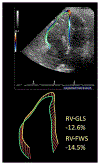
Utility of Left and Right Ventricular Strain in Arrhythmogenic Right Ventricular Cardiomyopathy: A Prospective Multicenter Registry
- PMID: 38113321
- PMCID: PMC10803132
- DOI: 10.1161/CIRCIMAGING.123.015671
Utility of Left and Right Ventricular Strain in Arrhythmogenic Right Ventricular Cardiomyopathy: A Prospective Multicenter Registry
Abstract
Background: Imaging evaluation of arrhythmogenic right ventricular cardiomyopathy (ARVC) remains challenging. Myocardial strain assessment by echocardiography is an increasingly utilized technique for detecting subclinical left ventricular (LV) and right ventricular (RV) dysfunction. We aimed to evaluate the diagnostic and prognostic utility of LV and RV strain in ARVC.
Methods: Patients with suspected ARVC (n = 109) from a multicenter registry were clinically phenotyped using the 2010 ARVC Revised Task Force Criteria and underwent baseline strain echocardiography. Diagnostic performance of LV and RV strain was evaluated using the area under the receiver operating characteristic curve analysis against the 2010 ARVC Revised Task Force Criteria, and the prognostic value was assessed using the Kaplan-Meier analysis.
Results: Mean age was 45.3±14.7 years, and 48% of patients were female. Estimation of RV strain was feasible in 99/109 (91%), and LV strain was feasible in 85/109 (78%) patients. ARVC prevalence by 2010 ARVC Revised Task Force Criteria is 91/109 (83%) and 83/99 (84%) in those with RV strain measurements. RV global longitudinal strain and RV free wall strain had diagnostic area under the receiver operating characteristic curve of 0.76 and 0.77, respectively (both P<0.001; difference NS). Abnormal RV global longitudinal strain phenotype (RV global longitudinal strain > -17.9%) and RV free wall strain phenotype (RV free wall strain > -21.2%) were identified in 41/69 (59%) and 56/69 (81%) of subjects, respectively, who were not identified by conventional echocardiographic criteria but still met the overall 2010 ARVC Revised Task Force Criteria for ARVC. LV global longitudinal strain did not add diagnostic value but was prognostic for composite end points of death, heart transplantation, or ventricular arrhythmia (log-rank P=0.04).
Conclusions: In a prospective, multicenter registry of ARVC, RV strain assessment added diagnostic value to current echocardiographic criteria by identifying patients who are missed by current echocardiographic criteria yet still fulfill the diagnosis of ARVC. LV strain, by contrast, did not add incremental diagnostic value but was prognostic for identification of high-risk patients.
Keywords: arrhythmogenic right ventricular dysplasia; diagnosis; echocardiography; prognosis; ventricular dysfunction.
Conflict of interest statement
Figures


Similar articles
-
Long term CMR follow up of patients with right ventricular abnormality and clinically suspected arrhythmogenic right ventricular cardiomyopathy (ARVC).J Cardiovasc Magn Reson. 2019 Dec 12;21(1):76. doi: 10.1186/s12968-019-0581-0. J Cardiovasc Magn Reson. 2019. PMID: 31831077 Free PMC article.
-
Arrhythmogenic Right Ventricular Cardiomyopathy: The Importance of Biventricular Strain in Risk-Stratification.Am J Cardiol. 2025 Apr 15;241:61-68. doi: 10.1016/j.amjcard.2025.01.006. Epub 2025 Jan 11. Am J Cardiol. 2025. PMID: 39805356
-
Right Ventricular Strain Derived from Cardiac MRI Feature Tracking for the Diagnosis and Prognosis of Arrhythmogenic Right Ventricular Cardiomyopathy.Radiol Cardiothorac Imaging. 2024 Jun;6(3):e230292. doi: 10.1148/ryct.230292. Radiol Cardiothorac Imaging. 2024. PMID: 38842456 Free PMC article.
-
Assessment of right ventricular systolic function by tissue Doppler echocardiography.Dan Med J. 2012 Mar;59(3):B4409. Dan Med J. 2012. PMID: 22381093 Review.
-
Clinical diagnosis and management strategies in arrhythmogenic right ventricular cardiomyopathy.J Electrocardiol. 2000;33 Suppl:49-55. doi: 10.1054/jclc.2000.20323. J Electrocardiol. 2000. PMID: 11265736 Review.
Cited by
-
Echocardiographic Updates in the Assessment of Cardiomyopathy.Curr Cardiol Rep. 2025 Jan 22;27(1):34. doi: 10.1007/s11886-024-02159-7. Curr Cardiol Rep. 2025. PMID: 39841294 Free PMC article. Review.
-
Cardiovascular imaging research and innovation in 2023.Eur Heart J Imaging Methods Pract. 2024 Apr 18;2(1):qyae029. doi: 10.1093/ehjimp/qyae029. eCollection 2024 Jan. Eur Heart J Imaging Methods Pract. 2024. PMID: 39045198 Free PMC article. Review.
-
Strain Analysis for Early Detection of Fibrosis in Arrhythmogenic Cardiomyopathy: Insights from a Preliminary Study.J Clin Med. 2024 Dec 6;13(23):7436. doi: 10.3390/jcm13237436. J Clin Med. 2024. PMID: 39685894 Free PMC article.
-
Right Ventricular Strain by Echocardiography: Current Clinical Applications and Future Directions for Mechanics Assessment of the Forgotten Ventricle.J Pers Med. 2025 May 30;15(6):224. doi: 10.3390/jpm15060224. J Pers Med. 2025. PMID: 40559087 Free PMC article. Review.
References
-
- Marcus FI, McKenna WJ, Sherrill D, Basso C, Bauce B, Bluemke DA, Calkins H, Corrado D, Cox MG, Daubert JP, et al. Diagnosis of arrhythmogenic right ventricular cardiomyopathy/dysplasia: proposed modification of the task force criteria. Circulation. 2010;121:1533–1541. doi: 10.1161/CIRCULATIONAHA.108.840827 - DOI - PMC - PubMed
-
- Yoerger DM, Marcus F, Sherrill D, Calkins H, Towbin JA, Zareba W, Picard MH; Multidisciplinary Study of Right Ventricular Dysplasia Investigators. Echocardiographic findings in patients meeting task force criteria for arrhythmogenic right ventricular cardiomyopathy. J Am Coll Cardiol. 2005;45:860–865. doi: 10.1016/j.jacc.2004.10.070 - DOI - PubMed
-
- Haugaa KH, Basso C, Badano LP, Bucciarelli-Ducci C, Cardim N, Gaemperli O, Galderisi M, Habib G, Knuuti J, Lancellotti P, et al. Comprehensive multi-modality imaging approach in arrhythmogenic cardiomyopathy – an expert consensus document of the European Association of Cardiovascular Imaging. Eur Heart J. 2017;18:237–253. doi: 10.1093/ehjci/jew229 - DOI - PMC - PubMed
Publication types
MeSH terms
Grants and funding
LinkOut - more resources
Full Text Sources
Miscellaneous

