

Morphological and metabolic asymmetries of the thalamic subregions in temporal lobe epilepsy predict cognitive functions
- PMID: 38114641
- PMCID: PMC10730825
- DOI: 10.1038/s41598-023-49856-x
Morphological and metabolic asymmetries of the thalamic subregions in temporal lobe epilepsy predict cognitive functions
Abstract
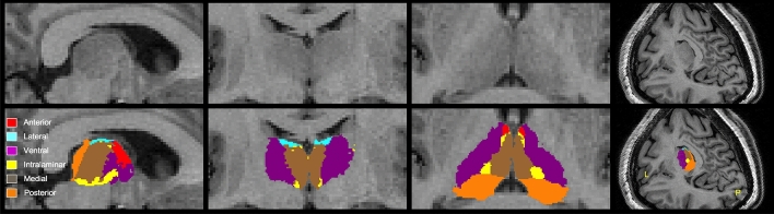
Both morphological and metabolic imaging were used to determine how asymmetrical changes of thalamic subregions are involved in cognition in temporal lobe epilepsy (TLE). We retrospectively recruited 24 left-TLE and 15 right-TLE patients. Six thalamic subnuclei were segmented by magnetic resonance imaging, and then co-registered onto Positron emission tomography images. We calculated the asymmetrical indexes of the volumes and normalized standard uptake value ratio (SUVR) of the entire and individual thalamic subnuclei. The SUVR of ipsilateral subnuclei were extensively and prominently decreased compared with the volume loss. The posterior and medial subnuclei had persistently lower SUVR in both TLE cases. Processing speed is the cognitive function most related to the metabolic asymmetry. It negatively correlated with the metabolic asymmetrical indexes of subregions in left-TLE, while positively correlated with the subnuclei volume asymmetrical indexes in right-TLE. Epilepsy duration negatively correlated with the volume asymmetry of most thalamic subregions in left-TLE and the SUVR asymmetry of ventral and intralaminar subnuclei in right-TLE. Preserved metabolic activity of contralateral thalamic subregions is the key to maintain the processing speed in both TLEs. R-TLE had relatively preserved volume of the ipsilateral thalamic volume, while L-TLE had relatively decline of volume and metabolism in posterior subnucleus.
© 2023. The Author(s).
Conflict of interest statement
The authors declare no competing interests.
Figures
References
MeSH terms
LinkOut - more resources
Full Text Sources
