

Icariin improves cardiac function and remodeling via the TGF-β1/Smad signaling pathway in rats following myocardial infarction
- PMID: 38115154
- PMCID: PMC10729580
- DOI: 10.1186/s40001-023-01588-4
Icariin improves cardiac function and remodeling via the TGF-β1/Smad signaling pathway in rats following myocardial infarction
Abstract
Background: Postinfarction cardiac remodeling presents a compensatory mechanism aimed at mitigating congestive heart failure. It is distinguished by progressive dilatation and hypertrophy of the ventricular chambers, fibrotic alterations, and prolonged apoptosis of cardiomyocytes. The primary objective of this study was to assess the effects of icariin on myocardial fibrosis and ventricular remodeling in rats subjected to myocardial infarction (MI).
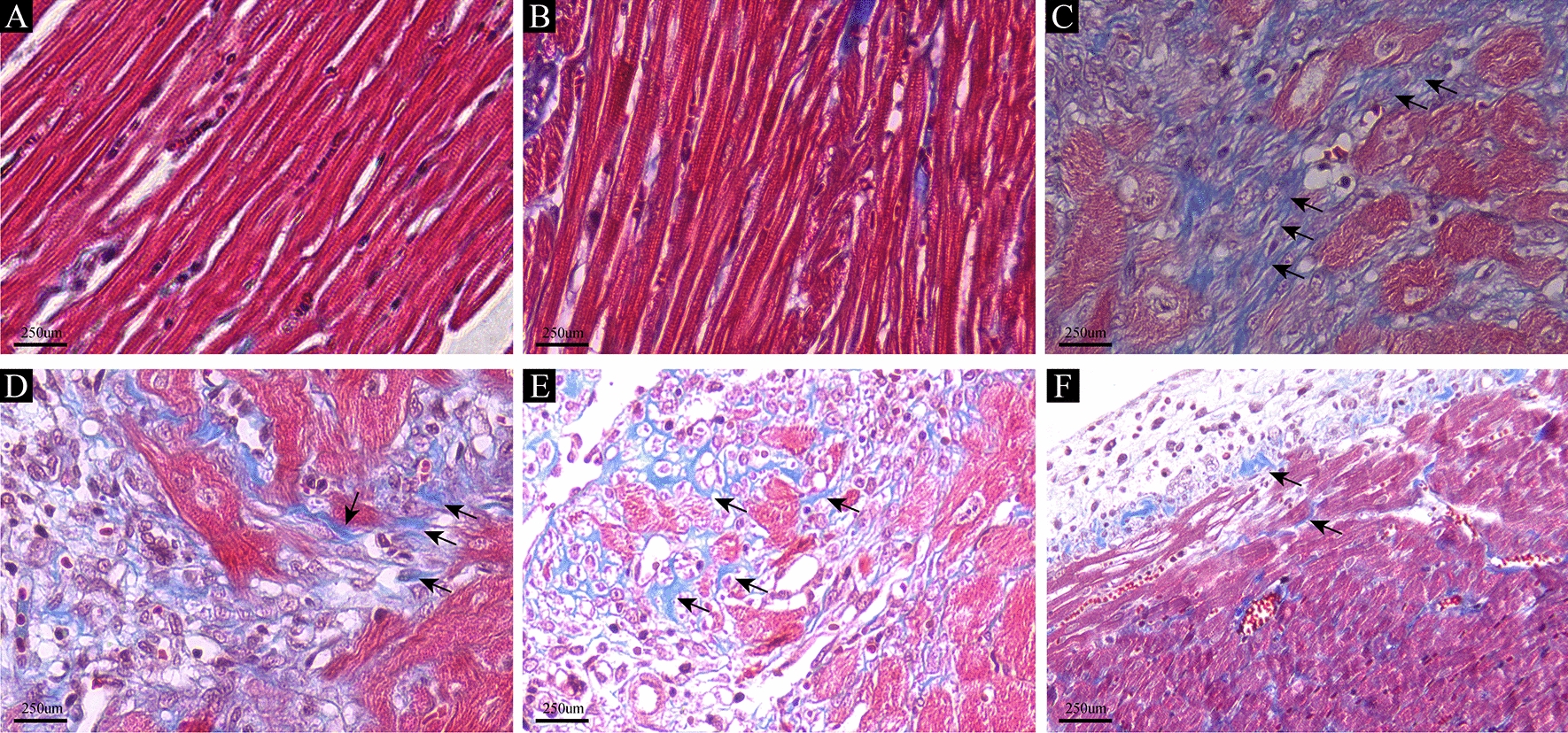
Methods: Male Sprague‒Dawley (SD) rats were subjected to randomization and subsequently divided into distinct groups: the control group, the sham group (undergoing sham operation), the MI group (experiencing ligation of the left anterior descending artery), and the icariin group. Within the icariin group, rats were further categorized into three different dose groups based on the administered icariin dosage: the MI30 group (30 mg/kg/day), the MI60 group (60 mg/kg/day), and the MI120 group (120 mg/kg/day). Cardiac function evaluation was carried out using echocardiography. Histological examinations, including hematoxylin and eosin (HE) staining, Masson staining, and immunohistochemistry studies, were conducted 90 days after the occurrence of MI. Additionally, Western blotting was employed to assess TGF-β1, p-Smad2, and p-Smad3 levels.
Results: The administration of icariin revealed a noteworthy enhancement in cardiac function among rats afflicted with left anterior descending coronary artery (LAD) ligation. In comparison to the icariin groups, the MI group exhibited reduced EF and FS, along with elevated LVEDD and LVESD. Furthermore, the cardiac fibrosis levels in the MI group rats exhibited a considerable increase compared to those in the icariin group. Notably, the levels of Collagen I, Collagen III, MMP2, and MMP9 were significantly higher in the MI group than in the icariin group, with evident distinctions. Moreover, the expression levels of TGF-β, IL-13, p-Smad2, and p-Smad3 were notably upregulated in the MI group compared to the icariin group.
Conclusions: In an experimental rat model of MI, the administration of icariin resulted in the amelioration of both cardiac function and remodeling processes, operating through the intricate TGF-β1/Smad signaling pathway.
Keywords: Icariin; Myocardial fibrosis; Myocardial infarction; TGF-β1/Smad signaling pathway; Ventricular remodeling.
© 2023. The Author(s).
Conflict of interest statement
The authors declare no competing interest.
Figures


Similar articles
-
Linggui Zhugan Decoction () Inhibits Ventricular Remodeling after Acute Myocardial Infarction in Mice by Suppressing TGF-β1/Smad Signaling Pathway.Chin J Integr Med. 2020 May;26(5):345-352. doi: 10.1007/s11655-018-3024-0. Epub 2019 Jan 9. Chin J Integr Med. 2020. PMID: 30623345
-
Zerumbone, a humulane sesquiterpene from Syringa pinnatifolia, attenuates cardiac fibrosis by inhibiting of the TGF-β1/Smad signaling pathway after myocardial infarction in mice.Phytomedicine. 2022 Jun;100:154078. doi: 10.1016/j.phymed.2022.154078. Epub 2022 Mar 31. Phytomedicine. 2022. PMID: 35405613
-
Soluble transforming growth factor-beta1 receptor II might inhibit transforming growth factor-beta-induced myofibroblast differentiation and improve ischemic cardiac function after myocardial infarction in rats.Coron Artery Dis. 2010 Sep;21(6):369-77. doi: 10.1097/MCA.0b013e32833ce0c3. Coron Artery Dis. 2010. PMID: 20613497
-
Qiliqiangxin Attenuates Cardiac Remodeling via Inhibition of TGF-β1/Smad3 and NF-κB Signaling Pathways in a Rat Model of Myocardial Infarction.Cell Physiol Biochem. 2018;45(5):1797-1806. doi: 10.1159/000487871. Epub 2018 Feb 28. Cell Physiol Biochem. 2018. PMID: 29510381
-
[Tea Polyphenols Regulate Renin-angiotensin-aldosterone System and Transforming Growth Factor-β1/Smads Signaling Pathway in Heart Failure Rats].Zhongguo Yi Xue Ke Xue Yuan Xue Bao. 2022 Jun;44(3):384-391. doi: 10.3881/j.issn.1000-503X.14385. Zhongguo Yi Xue Ke Xue Yuan Xue Bao. 2022. PMID: 35791933 Chinese.
Cited by
-
Icariin alleviates cisplatin-induced premature ovarian failure by inhibiting ferroptosis through activation of the Nrf2/ARE pathway.Sci Rep. 2024 Jul 27;14(1):17318. doi: 10.1038/s41598-024-67557-x. Sci Rep. 2024. PMID: 39068256 Free PMC article.
-
Exosomal miRNA-let-7i-5p from bone marrow mesenchymal stem cells protects against myocardial infarction by inhibiting myocardial apoptosis.Am J Transl Res. 2024 Nov 15;16(11):6528-6539. doi: 10.62347/VXND1945. eCollection 2024. Am J Transl Res. 2024. PMID: 39678588 Free PMC article.
-
Treatment of secondary uterine malignancy following radiotherapy for cervical cancer: a study based on the SEER database.BMC Womens Health. 2024 Aug 29;24(1):475. doi: 10.1186/s12905-024-03331-5. BMC Womens Health. 2024. PMID: 39210330 Free PMC article.
-
Traditional Chinese medicine compounds modulate signaling pathways to improve cardiac-related pathology.Front Pharmacol. 2025 Apr 2;16:1499060. doi: 10.3389/fphar.2025.1499060. eCollection 2025. Front Pharmacol. 2025. PMID: 40242436 Free PMC article. Review.
-
Advances in pharmacological research on myocardial remodeling agents: A decade in review.Medicine (Baltimore). 2025 Jun 6;104(23):e42757. doi: 10.1097/MD.0000000000042757. Medicine (Baltimore). 2025. PMID: 40489806 Free PMC article. Review.
References
-
- Park DY, An S, Attanasio S, Jolly N, Malhotra S, Doukky R, et al. Network meta-analysis comparing angiotensin receptor-neprilysin inhibitors, angiotensin receptor blockers, and angiotensin-converting enzyme inhibitors in heart failure with reduced ejection fraction. Am J Cardiol. 2023;187:84–92. doi: 10.1016/j.amjcard.2022.10.026. - DOI - PMC - PubMed
-
- Maizels L, Wasserstrum Y, Fishman B, Segev A, Ben-Nun D, Younis A, et al. Characterization of heart failure patients with reverse left ventricular remodeling postangiotensin receptor blockers/neprilysin inhibitors therapy. ESC Heart Fail. 2022;9(3):1682–1688. doi: 10.1002/ehf2.13801. - DOI - PMC - PubMed
MeSH terms
Substances
Grants and funding
- NO. 2022J0017/Science Research Foundation of Yunnan Provincial Department of Education Project
- NO. H-2017019/Medical Reserve Talent Training Program of Yunnan Provincial Health Commission of China
- NO. YNWR-MY-2020-024/Yunnan Provincial Program for the Cultivation of High-level Innovative Health Talent
LinkOut - more resources
Full Text Sources
Medical
Miscellaneous
