

Prenatal diagnosis of microcephaly through combined MRI and ultrasonography: Analysis of a case series
- PMID: 38115306
- PMCID: PMC10727632
- DOI: 10.1097/MD.0000000000036623
Prenatal diagnosis of microcephaly through combined MRI and ultrasonography: Analysis of a case series
Abstract
Introduction: Intrauterine microcephaly is a complex and lifelong condition that poses significant ethical challenges for clinicians and parents. The prognosis of microcephaly is highly variable and depends on the underlying cause and severity. In addition, microcephaly is often associated with various comorbidities, including intellectual disability, developmental delay, and epilepsy. Ultrasonography (US) is currently the most commonly used imaging modality for detecting microcephaly in the second trimester of pregnancy. However, antenatal brain magnetic resonance imaging (MRI) is increasingly being used as a more sensitive tool to identify structural abnormalities that may suggest a specific diagnosis. In this study, we report a case series of microcephaly diagnosed through the combination of MRI and US.
Patient concerns: How to utilize a combination of MRI and US to screen for fetal microcephaly.
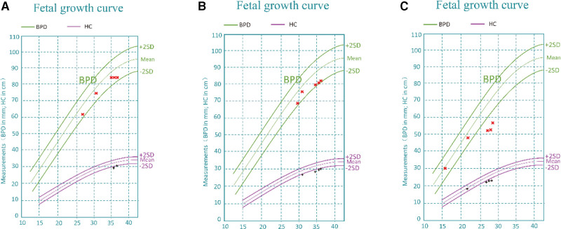
Diagnosis: Based on the results of US and MRI examinations, patient 1 was found to have other craniocerebral malformations, patient 2 demonstrated macrogyria, and patient 3 exhibited skull irregularities.
Interventions: The pregnancies of all 3 patients were terminated through the induction of labor by injecting Rivanol into the amniotic cavity.
Outcomes: The 3 patients were discharged after a period of observation.
Conclusion: US is an important tool for diagnosing fetal microcephaly. However, MRI can overcome the limitations of US and detect additional brain structural abnormalities, thereby providing more specific and valuable prenatal diagnostic information. Therefore, combining MRI and US has significant diagnostic value for fetal microcephaly.
Copyright © 2023 the Author(s). Published by Wolters Kluwer Health, Inc.
Conflict of interest statement
The authors have no funding and conflicts of interest to disclose.
Figures

References
-
- De Bie I, Boucoiran I. No. 380-investigation and management of prenatally identified microcephaly. J Obstet Gynaecol Can. 2019;41:855–61. - PubMed
-
- Chervenak FA, Jeanty P, Cantraine F, et al. The diagnosis of fetal microcephaly. Am J Obstet Gynecol. 1984;149:512–7. - PubMed
-
- Society for Maternal-Fetal Medicine (SMFM) Publications Committee. Ultrasound screening for fetal microcephaly following Zika virus exposure. Am J Obstet Gynecol. 2016;214:B2–4. - PubMed
-
- Cater SW, Boyd BK, Ghate SV. Abnormalities of the fetal central nervous system: prenatal US diagnosis with postnatal correlation. Radiographics. 2020;40:1458–72. - PubMed
-
- Salomon LJ, Alfirevic Z, Berghella V, et al. ISUOG practice guidelines (updated): performance of the routine mid-trimester fetal ultrasound scan. Ultrasound Obstet Gynecol. 2022;59:840–56. [Erratum in: Ultrasound Obstet Gynecol. 2022 Oct;60(4):591]. - PubMed
MeSH terms
LinkOut - more resources
Full Text Sources
Medical
