. 2023 Dec 19;4(6):e447.
doi: 10.1002/mco2.447. eCollection 2023 Dec.

NLRP3/IL-1β induced myeloid-derived suppressor cells recruitment and PD-L1 upregulation promotes oxaliplatin resistance of hepatocellular carcinoma

Affiliations

NLRP3/IL-1β induced myeloid-derived suppressor cells recruitment and PD-L1 upregulation promotes oxaliplatin resistance of hepatocellular carcinoma

Wenfeng Liu et al. MedComm (2020). .

Abstract

Oxaliplatin is commonly used as the first-line chemotherapeutic agent for advanced hepatocellular carcinoma (HCC). Unfortunately, the acquired resistance, limits the effectiveness of oxaliplatin and the underlying mechanisms remain unknown. Therefore, we explored the role of NOD-like receptor protein 3 (NLRP3)/IL-1β in mediating oxaliplatin resistance in HCC. We observed that NLRP3/IL-1β expression was much higher in oxaliplatin-resistant HCC cells. To further understand its impact on drug resistance, we knocked down NLRP3 and observed that it sensitized HCC cells to the growth-inhibitory effects of oxaliplatin and induced cell apoptosis. NLRP3/IL-1β overexpressing tumor cells also attracted polymorphonuclear myeloid-derived suppressor cells. Using mouse models, we demonstrated that NLRP3/IL-1β inhibition by short hairpin RNA or MCC950 effectively overcame oxaliplatin resistance. Furthermore, NLRP3/IL-1β inhibition resulted in reduced expression of PD-L1. We also found that PD-L1 antibody combined with NLRP3/IL-1β blockade displayed significant antitumor effect in HCC. Overall, our study provides compelling evidence supporting the essential role of NLRP3/IL-1β in conferring resistance to oxaliplatin and reshaping the immunosuppressive microenvironment in HCC. Targeting NLRP3/IL-1β presents a potential therapeutic target for overcoming oxaliplatin resistance and reshaping microenvironment of HCC.

Keywords: NLRP3/IL‐1β; PD‐L1; hepatocellular carcinoma; myeloid‐derived suppressor cells; oxaliplatin resistance.


Conflict of interest statement

The authors report no conflict of interest in this work.

Figures
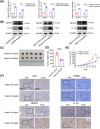
FIGURE 1
FIGURE 1
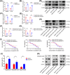
Oxaliplatin induced NLRP3 and IL‐1β expression in vitro and in vivo. (A and B) The mRNA and proteins levels of NLRP3 and IL‐1β in HCC cells treated with or without oxaliplatin. (C and D) The appearance and growth curves of the subcutaneous tumors. (E) Tumor weight was analyzed between different groups. (F) Representative pictures of IHC staining using Ki67 and TUNEL as markers of proliferation and apoptosis in the tumor tissues. *p < 0.05, **p < 0.01, ***p < 0.001.
FIGURE 2
FIGURE 2
NLRP3 was highly expressed in oxaliplatin‐resistant HCC cells and involved in oxaliplatin resistance. (A–C) The mRNA expression of NLRP3 and IL‐1β in HCC cells and oxaliplatin‐resistant HCC (Hep3B‐OXA, MHCC97H, and Hepa1‐6‐OXA) cells. (D) NLRP3 and IL‐1β proteins in HCC and oxaliplatin‐resistant HCC cells were detected. (E–G) Hep3B‐OXA, MHCC97H, and Hepa1‐6‐OXA cells were transfected with scramble shRNA and lentiviral shRNA targeting NLRP3. RT‐PCR confirmed the downregulation of NLRP3 and L‐1β. (H) NLRP3 and IL‐1β proteins in oxaliplatin‐resistant HCC cells after NLRP3 silencing. (I) The IC50 values of oxaliplatin‐resistant HCC cells transfected with NLRP3 shRNA were significantly lower than those transfected with control lentiviral vectors. (J) The proapoptotic effect of NLRP3 silencing on oxaliplatin‐induced apoptosis was confirmed by flow cytometry in oxaliplatin‐resistant HCC cells. (K) Cleaved‐cas3, bcl2, and bax proteins were examined in different group. *p < 0.05, **p < 0.01, ***p < 0.001.
FIGURE 3
FIGURE 3
NLRP3/IL‐1β signaling drove PMN‐MDSCs recruitment and affected PD‐L1 expression. (A) The IL‐1β protein in conditional media (CM) from Hep3B, Hep3B‐OXA, Hep3B‐OXA‐control, and Hep3B‐OXA‐shNLRP3 was detected by ELISA. (B) The IL‐1β protein in CM from MHCC97H, MHCC97H‐OXA, MHCC97H‐OXA‐control, and MHCC97H‐OXA‐shNLRP3. (C) The IL‐1β protein in CM from Hepa1‐6, Hepa1‐6‐OXA, Hepa1‐6‐OXA‐control, and Hepa1‐6‐OXA‐shNLRP3. (D and E) MDSCs migration assays using CM from different cells. (F) The overall survival rate in HCC patients with high (n = 180) and low IL‐1β level (n = 179) using data from TCGA dataset. (G and H) Positive correlations between NLRP3/IL‐1β and CD274 (PD‐L1) using data from TCGA dataset. (I) PD‐L1 mRNA levels were significantly lower in oxaliplatin‐resistant cells transfected with NLRP3 shRNA. (J) PD‐L1 membrane expressions were downregulated after NLRP3 knockdown detected by flow cytometry. (K) PD‐L1 protein was detected in cells transfected with NLRP3 shRNA. *p < 0.05, **p < 0.01, ***p < 0.001.
FIGURE 4
FIGURE 4
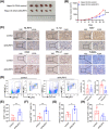
NLRP3/IL‐1β facilitated oxaliplatin‐resistant HCC progression in vivo. (A) The appearance of the subcutaneous tumors from Hepa1‐6‐OXA‐control and Hepa1‐6‐OXA‐shNLRP3 group. (B) Tumor growth curves of each group. (C) IHC staining of NLRP3, IL‐1β, Ki67, CD8, PD‐L1, and CD11b. (D) There was a significant reduction in CD11b+Ly6G+ PMN‐MDSCs level in the Hepa1‐6‐OXA‐shNLRP3 group, while the changes of CD11b+Ly6C+ M‐MDSCs showed no statistical significance. (E) The CD3+CD8+ T cells was enriched in the Hepa1‐6‐OXA‐shNLRP3 group. (F–H) The granzyme B, Perforin, and CTLA4 positive CD8+ T cells are depicted. *p < 0.05, **p < 0.01, ***p < 0.001.
FIGURE 5
FIGURE 5
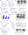
MCC950 affected PMN‐MDSC recruitment and inhibited PD‐L1 expression. (A and B) The mRNA expression of NLRP3 and IL‐1β was reduced in oxaliplatin‐resistant HCC (Hep3B‐OXA, MHCC97H, and Hepa1‐6‐OXA) cells treated with MCC950. (C) NLRP3 and IL‐1β proteins were lower in oxaliplatin‐resistant cells treated with MCC950. (D) The IC50 values of oxaliplatin‐resistant HCC cells treated with MCC950 were significantly lower than those treated with PBS. (E) The proapoptotic effect of MCC950 on oxaliplatin‐induced apoptosis was confirmed by flow cytometry in oxaliplatin‐resistant HCC cells. (F) Cleaved‐cas3, bcl2, and bax proteins in indicated cells exposed to MCC950 or PBS. (G) The IL‐1β protein in CM from indicated cells treated with MCC950 or PBS was detected by ELISA. (H) MDSC migration ability was tested utilizing CM from Hepa1‐6‐OXA cells exposed to MCC950 or PBS. (I) PD‐L1 mRNA was reduced in indicated cells treated with MCC950. (J) The PD‐L1 protein was lower in indicated treatment groups. *p < 0.05, **p < 0.01, ***p < 0.001.
FIGURE 6
FIGURE 6
IL‐1β blocking inhibited PMN‐MDSCs recruitment and enhanced CD8+T cell‐killing ability. (A and B) The tumor appearance and growth curves were shown in different group. (C) The infiltration of CD3+CD8+ T cells was much higher in mice treated with MCC950 or anti‐IL‐1β. (D) A significant reduction of CD11b+Ly6G+ PMN‐MDSCs infiltration when treated with MCC950 or IL‐1β antibody, while the changes in CD11b+Ly6C+ M‐MDSCs showed no statistical significance. *p < 0.05, **p < 0.01, ***p < 0.001.
FIGURE 7
FIGURE 7
IL‐1β blocking enhanced the effect of anti‐PD‐L1 therapy. (A and B) The tumor appearance and growth curves were shown in different group. (C–G) The proportions of MDSCs, CD8+ T cells, along with the levels of granzyme B, Perforin, and CTLA4 positive CD8+ T cells are displayed. *p < 0.05, **p < 0.01, ***p < 0.001.

References

    1. Xia C, Dong X, Li H, et al. Cancer statistics in China and United States, 2022: profiles, trends, and determinants. Chin Med J (Engl). 2022;135(5):584‐590. - PMC - PubMed
    1. Peng H, Zhu E, Zhang Y. Advances of cancer‐associated fibroblasts in liver cancer. Biomark Res. 2022;10(1):59. - PMC - PubMed
    1. Couri T, Pillai A. Goals and targets for personalized therapy for HCC. Hepatol Int. 2019;13(2):125‐137. - PubMed
    1. Raoul JL, Forner A, Bolondi L, et al. Updated use of TACE for hepatocellular carcinoma treatment: how and when to use it based on clinical evidence. Cancer Treat Rev. 2019;72:28‐36. - PubMed
    1. Guerrero‐Juarez CF, Lee GH, Liu Y, et al. Single‐cell analysis of human basal cell carcinoma reveals novel regulators of tumor growth and the tumor microenvironment. Sci Adv. 2022;8(23):eabm7981. - PMC - PubMed