. 2023 Dec 1;64(15):25.
doi: 10.1167/iovs.64.15.25.

Mechanical Disruption of the Inner Limiting Membrane In Vivo Enhances Targeting to the Inner Retina

Affiliations

Mechanical Disruption of the Inner Limiting Membrane In Vivo Enhances Targeting to the Inner Retina

Jiun L Do et al. Invest Ophthalmol Vis Sci. .

Abstract

Purpose: To evaluate the effects of mechanical disruption of the inner limiting membrane (ILM) on the ability to target interventions to the inner neurosensory retina in a rodent model. Our study used an animal model to gain insight into the normal physiology of the ILM and advances our understanding of the effects of mechanical ILM removal on the viral transduction of retinal ganglion cells and retinal ganglion cell transplantation.

Methods: The ILM in the in vivo rat eye was disrupted using mechanical forces applied to the vitreoretinal interface. Immunohistology and electron microscopy were used to verify the removal of the ILM in retina flatmounts and sections. To assess the degree to which ILM disruption enhanced transvitreal access to the retina, in vivo studies involving intravitreal injections of adeno-associated virus (AAV) to transduce retinal ganglion cells (RGCs) and ex vivo studies involving co-culture of human stem cell-derived RGCs (hRGCs) on retinal explants were performed. RGC transduction efficiency and transplanted hRGC integration with retinal explants were evaluated by immunohistology of the retinas.

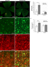
Results: Mechanical disruption of the ILM in the rodent eye was sufficient to remove the ILM from targeted retinal areas while preserving the underlying retinal nerve fiber layer and RGCs. Removal of the ILM enhanced the transduction efficiency of intravitreally delivered AAV threefold (1380.0 ± 290.1 vs. 442.0 ± 249.3 cells/mm2; N = 6; P = 0.034). Removal of the ILM was also sufficient to promote integration of transplanted RGCs within the inner retina.

Conclusions: The ILM is a barrier to transvitreally delivered agents including viral vectors and cells. Mechanical removal of the ILM is sufficient to enhance access to the inner retina, improve viral transduction efficiencies of RGCs, and enhance cellular integration of transplanted RGCs with the retina.


Conflict of interest statement

Disclosure: J.L. Do, Auxilium Biotechnologies (C), Nicox (C), VoxelCloud (C); N. Pedroarena-Leal, None; M. Louie, None; P. Avila Garcia, None; A. Alnihmy, None; A. Patel, None; R.N. Weinreb, Abbvie (C), Alcon (C), Allergan (C), Amydis (C), Centervue (R), Editas (C), Eyenovia (C), Heidelberg Engineering (R), Iantrek (C), Implandata (C), IOPtic (C), Konan (R), Nicox (C), Optovue (R), Santen (C), Topcon (C), Toromedes (F, P), Zeiss (P, R), Zilia (R); K.J. Wahlin, None; A. La Torre Vila, None; D.S. Welsbie, Perceive Biotherapeutics (C, F)

Figures

Figure 1.
Figure 1.
Mechanical disruption of the vitreoretinal interface removes the ILM in the rat eye. (A) High-magnification image of a micro-serrated nitinol loop used in human vitreoretinal surgery to create breaks in the ILM. (B) Intraoperative image of a micro-serrated nitinol loop being applied to the vitreoretinal surface to remove the ILM in a rat eye. (C) Representative image of a retinal flatmount stained for laminin immediately following ILM removal and magnified inset demonstrating small breaks (white arrowheads) in the ILM with conservative treatment. (D) Moderate treatment of the ILM creates more contiguous tears (inset, white arrowheads) in the ILM. (E) Heavy treatment can cause larger breaks (inset, white arrowheads) in the ILM. (F) Electron microscopy of the retina with an intact ILM demonstrated the presence of the ILM and Müller glia endfeet (black arrows) overlying axon bundles (blue asterisks) and cell nuclei (red asterisks). (G) In contrast, in retinas treated with mechanical disruption to remove the ILM, the ILM is absent while axon bundles (blue asterisks) are preserved. (H) Two weeks after removal of the ILM, breaks in the ILM (inset, white arrowheads) were still present. Scale bars: 2000 µm (CE, H); 400 µm (insets, CE, H); 5 µm (F, G).
Figure 2.
Figure 2.
Inner retinal cellular organization is maintained following mechanical removal of the ILM in the rat eye. Representative cross-sectional images of retinas without and with removal of the ILM from rats that ubiquitously express GFP. (A) Naïve animals express GFP in all layers of the retina. (B, C) Immunolabeling for laminin using two different antibodies demonstrated a continuous layer of immunopositivity along the inner retinal surface. (D) Nuclear staining with 4′,6-diamidino-2-phenylindole (DAPI) highlights the ganglion cell (*), bipolar (+), and photoreceptor (#) cell layers. (E) Merged image demonstrating the retinal histology with an intact ILM. (F) Mechanical removal of the ILM in retinas from GFP-expressing rats does not distort the gross anatomy of the retina. (G, H) Two different antibodies that immunolabel laminin demonstrated fragmentation of the ILM (white arrowheads) following mechanical treatment. (I) DAPI staining of cell nuclei demonstrated retention of all cellular layers (ganglion cell [*], bipolar [+], and photoreceptor [#] cell layers) in the setting of ILM removal. (J) Merged image of immunolabeled retina following ILM removal demonstrating successful removal of the ILM with preservation of the inner retinal structures. (K) Quantification of RGC survival based on the density of SNCG immunolabeled cells demonstrated similar RGC densities in retinas with intact ILMs and following ILM removal in focal areas where the ILM was removed and over the entire retina. (L) Quantification of RGC SNCG intensity was also similar among groups, suggestive of comparable cell viabilities. Error bars represent SE; n = 6 animals per group. Scale bars: 500 µm.
Figure 3.
Figure 3.
Transient glial activation occurs in the retina following mechanical removal of the ILM in the rat eye. Representative images of immunolabeled inflammatory cells in retinal flatmounts from eyes in which the ILM was left intact or mechanically disrupted. (AC) In retinas with an intact ILM, CD11b-positive glial cells were loosely distributed along the retinal vasculature and adopted ramified morphologies. (DF) One day after mechanical disruption of the ILM, there was an increase in CD11b-positive immunolabeling in areas where the ILM had been disrupted (as demonstrated by the lack of laminin immunolabeling), and immunolabeled glial cells could be seen adopting ameboid morphologies. (GI) By day 3 after ILM disruption, CD11b-positive immunolabeling in areas of ILM disruption was significantly reduced, and clustering of the glial cells was no longer obvious. (JL) Five days after ILM disruption, a majority of CD11b-positive cells within the area of ILM disruption had reverted back to assuming ramified morphologies. Scale bar: 100 µm.
Figure 4.
Figure 4.
Viral transduction of RGCs by intravitreal injection is enhanced by mechanical removal of the ILM. Representative images of retinal flatmounts from eyes that received an intravitreal injection of GFP-expressing AAV2 with an intact ILM (A) or mechanical treatment to disrupt the ILM (E). Immunolabeling demonstrated that a greater number of RGCs transduced to express GFP in an eye in which the ILM is disrupted (F) compared to an eye in which the ILM is intact (B). Immunolabeling for SNCG demonstrated similar SNCG-positive RGC densities in an eye with an intact ILM (C) and with the ILM removed (G). Merged image from an eye with an intact ILM (D) and with the ILM removed (H) demonstrating colocalization of GFP and SNCG expression. (I) Quantification of GFP-positive cell densities demonstrated a greater number of transduced RGCs densities in retinas following ILM removal compared to retinas with intact ILMs in focal areas where the ILM was removed and over the entire retina. (J) Quantification of RGC GFP intensity was not statistically significant between retinas with intact ILMs and following ILM removal, although there was a trend toward greater GFP intensity in focal areas where the ILM was removed. *P < 0.05, Student's t-test. Error bars represent SE, n = 6 animals per group. Scale bars: 1000 µm (A, E); 50 µm (BD, FH).
Figure 5.
Figure 5.
Mechanical removal of the ILM allows transplanted RGCs to integrate into the inner retina. (A) Confocal image of a retinal explant with an intact ILM demonstrated by laminin immunolabeling (blue) cocultured with human stem cell-derived RGCs that express tdTomato (red). Transplanted RGCs are observed on top of the ILM without penetration into the inner retina or host ganglion cell layer. (B) Confocal image of a retinal explant in which the ILM has been removed cocultured with hRGCs (red). Transplanted RGCs can be observed in the ganglion cell layer and extend process (white arrowhead) in the inner plexiform layer and bipolar cell layer. (C) Three-dimensional reconstruction from confocal images of a retinal explant in which the ILM has been removed and hRGCs have been cultured on the inner retinal surface. Transplanted RGC-derived processes (white arrowheads) are shown extending into outer retinal layers. This was not observed with retinal explants in which the ILM was left intact (not shown).

References

    1. Inoue T, Hosokawa M, Morigiwa K, Ohashi Y, Fukuda Y. Bcl-2 overexpression does not enhance in vivo axonal regeneration of retinal ganglion cells after peripheral nerve transplantation in adult mice. Journal of Neuroscience. 2002; 22: 4468–4477. - PMC - PubMed
    1. Weinreb RN, Aung T, Medeiros FA. The pathophysiology and treatment of glaucoma: a review. JAMA. 2014; 311: 1901–1911. - PMC - PubMed
    1. Sluch VM, Chamling X, Liu MM, et al. .. Enhanced stem cell differentiation and immunopurification of genome engineered human retinal ganglion cells. Stem Cells Transl Med. 2017; 6: 1972–1986. - PMC - PubMed
    1. Sluch VM, Davis CO, Ranganathan V, et al. .. Differentiation of human ESCs to retinal ganglion cells using a CRISPR engineered reporter cell line. Sci Rep. 2015; 5: 1–17. - PMC - PubMed
    1. Vergara MN, Flores-Bellver M, Aparicio-Domingo S, et al. .. Three-dimensional automated reporter quantification (3D-ARQ) technology enables quantitative screening in retinal organoids. Development. 2017; 144: 3698–3705. - PMC - PubMed

Publication types