. 2024 Jan 23;43(1):113596.
doi: 10.1016/j.celrep.2023.113596. Epub 2023 Dec 20.

The HLA-II immunopeptidome of SARS-CoV-2

Affiliations

The HLA-II immunopeptidome of SARS-CoV-2

Shira Weingarten-Gabbay et al. Cell Rep. .

Abstract

Targeted synthetic vaccines have the potential to transform our response to viral outbreaks, yet the design of these vaccines requires a comprehensive knowledge of viral immunogens. Here, we report severe acute respiratory syndrome coronavirus 2 (SARS-CoV-2) peptides that are naturally processed and loaded onto human leukocyte antigen-II (HLA-II) complexes in infected cells. We identify over 500 unique viral peptides from canonical proteins as well as from overlapping internal open reading frames. Most HLA-II peptides colocalize with known CD4+ T cell epitopes in coronavirus disease 2019 patients, including 2 reported immunodominant regions in the SARS-CoV-2 membrane protein. Overall, our analyses show that HLA-I and HLA-II pathways target distinct viral proteins, with the structural proteins accounting for most of the HLA-II peptidome and nonstructural and noncanonical proteins accounting for the majority of the HLA-I peptidome. These findings highlight the need for a vaccine design that incorporates multiple viral elements harboring CD4+ and CD8+ T cell epitopes to maximize vaccine effectiveness.

Keywords: CD4(+) T cell; CP: Immunology; CP: Microbiology; HLA-II; SARS-CoV-2; antigen processing and presentation; immunity; immunopeptidome; noncanonical protein; viral antigen.


Conflict of interest statement

Declaration of interests S.W.-G., D.-Y.C., S.S., K.R.C., N.H., S.A.C., J.G.A., M.S., and P.C.S. are named co-inventors on a patent application related to this work, filed by The Broad Institute, that is being made available in accordance with the COVID-19 technology licensing framework to maximize access to university innovations. N.H. is a founder of Neon Therapeutics (now BioNTech US), was a member of its scientific advisory board, and holds shares. N.H. is also an advisor for IFM Therapeutics. S.A.C. is a member of the scientific advisory boards of Kymera, PTM BioLabs, Seer, and PrognomIQ. J.G.A. is a past employee of Neon Therapeutics (now BioNTech US). P.C.S. is a cofounder of and consultant to Sherlock Biosciences and Delve Biosciences and a board member of Danaher Corporation and holds equity in the companies.

Figures

Figure 1.
Figure 1.. HLA-II immunopeptidome profiling of SARS-CoV-2-infected cells
(A) Schematic of the experimental workflow. (B) HLA-II expression measured by flow cytometry using a fluorescein isothiocyanate (FITC)-conjugated anti HLA-DR/DP/DQ antibody. Gating was based on unstained cells. (C) SARS-CoV-2 infection levels in A549/AT and HEK293T/AT cells with and without CIITA transduction. The infection was quantified using immunofluorescence staining for the nucleocapsid protein. Bar height is equal to the mean percentage of infected cells counted in 12 different fields; error bars correspond to mean ± SD. (D) Length distribution of the eluted peptides in SARS-CoV-2-infected cells shown for peptides that were derived from human proteins (black), SARS-CoV-2 proteins (blue), and BSA (gray). (E) GibbsCluster deconvoluted motifs of the eluted peptides. (F) The percentage of peptides originating from bovine proteins detected in the HLA-I and HLA-II immunopeptidomes. HLA-I data are from Weingarten-Gabbay et al. (G) Length distribution of human and bovine peptides in the A549 and HEK293T HLA-I and the HLA-II immunopeptidomes. See also Figure S1 and Table S1.
Figure 2.
Figure 2.. SARS-CoV-2 HLA-II immunopeptidome
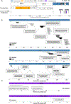
(A) Summary of viral proteins presented on HLA-II complexes in infected A549/ATC and HEK293T/ATC cells. (B) The location of HLA-II peptides in canonical structural proteins M, N, and S. Peptides detected in A549/ATC and HEK293T/AT/C cells are depicted in black and gray, respectively. (C) The location of HLA-II peptides in 2 noncanonical internal ORFs: ORF9b and ORF3c. See also Figures S2 and S3.
Figure 3.
Figure 3.. Systematic comparison between the SARS-CoV-2 HLA-II peptides and reported CD4+ T cell epitopes
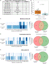
(A) A list of 9 studies that identified CD4+ T cells epitopes in COVID-19 patients as described in Table 1 of Grifoni et al. Studies that included peptides from all canonical SARS-CoV-2 proteins are highlighted with an asterisk. (B) Comparing the immunogenicity of SARS-CoV-2 proteins that were detected on the HLA-II complex to proteins that were not. For each protein (each dot), we plotted the fraction of CD4+ T cell epitopes from all of the detected epitopes in 4 genome-wide studies (denoted with asterisk in A). The box shows the quartiles, the bar indicates median, and the whiskers show the distribution. Wilcoxon rank-sum p value is shown. (C–E) Comparing the location of HLA-II peptides that were detected by LC-MS/MS in infected A549/AT/CIITA and HEK293T/AT/CIITA cells to previously reported CD4+ T cell epitopes. (Left) Heatmap showing the density of HLA-II peptides and the reported CD4+ T cell epitopes across individual viral proteins. Rows were normalized according to the maximal density in each row. (Right) Venn diagram showing the number of amino acids that are covered by HLA-II peptides, CD4+ T cell epitopes, and both. To reduce background levels, we counted amino acids (aa) with greater coverage than the mean of each row. Hypergeometric p value is shown for each viral protein.
Figure 4.
Figure 4.. SARS-CoV-2 protein representation on the HLA-I and HLA-II complexes
(A) A bar chart showing the number of HLA-II peptides detected in SARS-CoV-2-infected A549/AT/CIITA and HEK293T/AT/CIITA cells for each viral protein. Inside the frame is a pie chart showing the relative abundance of peptides derived from structural proteins, nonstructural proteins, accessory proteins, and noncanonical ORFs. (B) Similar to (A) for HLA-I peptides reported in Weingarten-Gabbay et al.. (C) The source proteins for the HLA-II processing and presentation pathway. Viruses are endocytosed by an APC. The viral structural proteins are cleaved within the endosomal-lysosomal compartment and loaded onto HLA-II complexes. (D) The source proteins for the HLA-I processing and presentation pathway. Viral proteins are produced from the translation of genomic and subgenomic viral RNAs in infected cells. These proteins are cleaved and loaded onto HLA-I complexes.

Update of

References

    1. Wherry EJ, and Barouch DH (2022). T cell immunity to COVID-19 vaccines. Science 377, 821–822. - PubMed
    1. Sette A, and Crotty S (2021). Adaptive immunity to SARS-CoV-2 and COVID-19. Cell 184, 861–880. - PMC - PubMed
    1. Pardi N, Hogan MJ, Porter FW, and Weissman D (2018). mRNA vaccines — a new era in vaccinology. Nat. Rev. Drug Discov. 17, 261–279. - PMC - PubMed
    1. Neale I, Ali M, Kronsteiner B, Longet S, Abraham P, Deeks AS, Brown A, Moore SC, Stafford L, Dobson SL, et al. (2023). CD4+ and CD8+ T Cell and Antibody Correlates of Protection against Delta Vaccine Breakthrough Infection: A Nested Case-Control Study within the PITCH Study. Preprint at medRxiv. - PMC - PubMed
    1. Sahin U, Muik A, Vogler I, Derhovanessian E, Kranz LM, Vormehr M, Quandt J, Bidmon N, Ulges A, Baum A, et al. (2021). BNT162b2 vaccine induces neutralizing antibodies and poly-specific T cells in humans. Nature 595, 572–577. - PubMed