. 2023 Dec 22;9(51):eadj1397.
doi: 10.1126/sciadv.adj1397. Epub 2023 Dec 20.

NET formation is a default epigenetic program controlled by PAD4 in apoptotic neutrophils

Affiliations

NET formation is a default epigenetic program controlled by PAD4 in apoptotic neutrophils

Yanfang Peipei Zhu et al. Sci Adv. .

Abstract

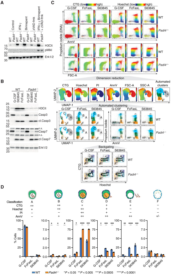
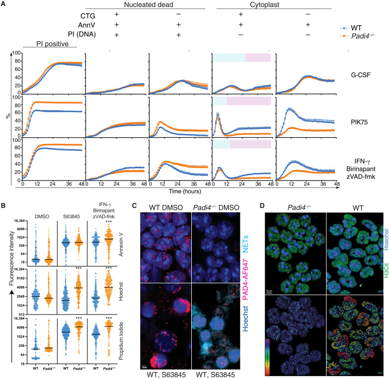
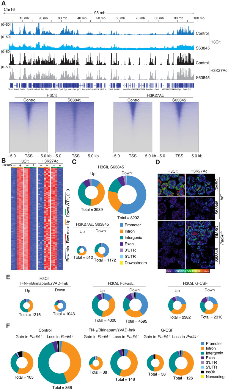
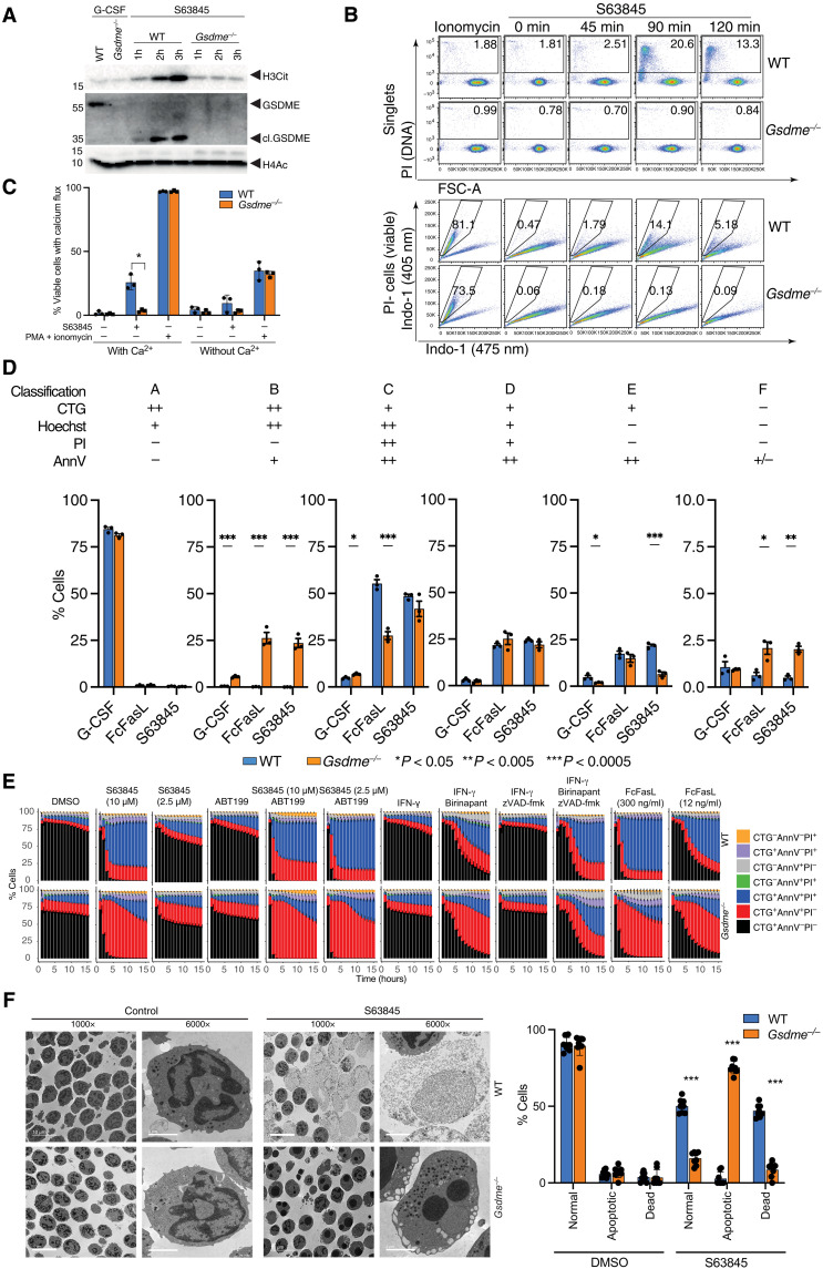
Neutrophil extracellular traps (NETs) not only counteract bacterial and fungal pathogens but can also promote thrombosis, autoimmunity, and sterile inflammation. The presence of citrullinated histones, generated by the peptidylarginine deiminase 4 (PAD4), is synonymous with NETosis and is considered independent of apoptosis. Mitochondrial- and death receptor-mediated apoptosis promote gasdermin E (GSDME)-dependent calcium mobilization and membrane permeabilization leading to histone H3 citrullination (H3Cit), nuclear DNA extrusion, and cytoplast formation. H3Cit is concentrated at the promoter in bone marrow neutrophils and redistributes in a coordinated process from promoter to intergenic and intronic regions during apoptosis. Loss of GSDME prevents nuclear and plasma membrane disruption of apoptotic neutrophils but prolongs early apoptosis-induced cellular changes to the chromatin and cytoplasmic granules. Apoptotic signaling engages PAD4 in neutrophils, establishing a cellular state that is primed for NETosis, but that occurs only upon membrane disruption by GSDME, thereby redefining the end of life for neutrophils.


Figures

Fig. 1.
Fig. 1.. PAD4 is activated by apoptotic and necroptotic stimuli and controls cytoplast formation.
(A and B) Protein expression levels of H3Cit, pMLKL, (cl.) caspase 3, and Erk1/2 in freshly isolated WT and Padi4−/− BM neutrophils stimulated with apoptotic and necroptotic stimuli. (C) Flow cytometry evaluation of cell size (FSC-A) and granularity (SSC-A), propidium iodide (PI), annexin V (AnnV), CellTracker Green (CTG), and Hoechst in WT and Padi4−/− neutrophils stimulated as indicated. (D) Statistical analysis of the proportion of each cell cluster identified by dimension reduction (UMAP) and automated clustering (FLOWSOM) from (C). Means ± SEM, n = 3 in each stimuli group, *P < 0.05, **P < 0.005, ***P < 0.0005, ****P < 0.0001, WT versus Padi4−/−, unpaired t test. n.s., not significant.
Fig. 2.
Fig. 2.. PAD4 mediates cytoplast formation after disruption of plasma membrane integrity.
(A) Kinetic changes in WT and Padi4−/− neutrophil viability in 48-hour live-cell imaging in the presence of G-CSF (100 ng/ml), 1 μM PIK75, or combinations of 2 μM BPT, 10 μM zVAD-fmk, and IFN-γ (100 ng/ml). Data represent mean and SEM of independent biological samples from five WT and seven Padi4−/− mice. Blue color in column 4 represents dying neutrophils. Pink color in column 4 represents dead neutrophils lacking a nucleus (cytoplast). (B) Quantitative fluorescence intensity of WT and Padi4−/− neutrophils at 12 hours classified according to staining with Hoechst, CTG, PI, and AnnV. ***P < 0.0001, WT versus Padi4−/−. (C) Fluorescence microscopy showing externalized DNA from neutrophils treated with S63845. Neutrophils were stained with Hoechst and a PAD4 antibody coupled to AF647. A 3D nuclear mask was generated to identify DNA outside the nuclear outline to separate intranuclear (dark blue) from extranuclear (aqua) DNA (NETs). (D) Super-resolution microscopy of H3Cit distribution in WT and Padi4−/− neutrophils indicating the difference in intensity and distribution of signal. A bounding box metric indicates the continuity of H3Cit signal by assessing the length of the longest principal axis inside a rectangular cuboid object and whose local axes are aligned along the principal axis of the object. Data are representative of three independent experiments.
Fig. 3.
Fig. 3.. Cell death–triggered epigenetic changes in dying neutrophils.
(A) Top: Genome tracks of H3Cit and H3K27Ac of WT and Padi4−/− control neutrophils or treated with S63845 and analyzed using CUT&Tag. Data representative to three biological replicates. Bottom: Heatmap representation of WT neutrophil H3Cit and H3K27Ac at the TSS following S63845 treatment for 2.5 hours. (B) Heatmap representation of unique differential H3Cit or H3K27Ac peaks at specific genomic loci using CUT&Tag in independent samples of neutrophils treated with S63845 for 2.5 hours or controls. (C) Genomic feature analysis of H3Cit and H3K27Ac differential peaks in WT neutrophils treated with S63845 and analyzed by CUT&Tag. (D) Super-resolution microscopy of H3Cit and H3K27Ac distribution in WT and Padi4−/− neutrophils treated with S63845 and controls. A bounding box metric indicates the continuity of signal. (E) Genomic feature analysis of H3Cit CUT&Tag signal in WT neutrophils treated with S63845, IFN-γ + birinapant + zVAD-fmk, FcFasL, or G-CSF. (F) ATAC-seq evaluation of differential chromatin accessibility in freshly isolated WT and Padi4−/− control neutrophils or treated with G-CSF or IFN-γ + birinapant + zVAD-fmk. 3′UTR, 3′ untranslated region.
Fig. 4.
Fig. 4.. Mcl-1 antagonism triggers GSDME cleavage leading to Ca2+ influx, PAD4 activation, and H3Cit accumulation.
(A) Protein expression levels of H3Cit, (cl.) GSDME, (cl.) caspase 3, and H4Ac in freshly isolated WT and Gsdme−/− BM neutrophils or treated with S63845 and G-CSF. (B) Top: Flow cytometry evaluation of PI+ cells in single WT and Gsdme−/− neutrophils treated with S63845 for 0, 45, 90, and 120 min. Frequency of PI+ cells are indicated on each plot. Bottom: Ca2+ influx is determined in PI-(viable) neutrophils by switch of Indo-1 (475 nm)–positive to Indo-1 (405 nm)–positive staining. Frequency of Indo-1 (405 nm) calcium fluxing cells is indicated on each plot. (C) Calcium flux in neutrophils treated with S63845 for 90 min or PMA + ionomycin for 30 s in the presence or absence of 1.8 mM extracellular calcium in Dulbecco’s modified Eagle’s medium (DMEM). (D) Statistical analysis of automated cell clusters identified by flow cytometry measurement of cell size and granularity, PI, AnnV, CTG, and Hoechst in WT and Gsdme−/− neutrophils treated as indicated. Means ± SEM, n = 3 independent biological replicates, significance is determined by unpaired t tests, *P < 0.05, **P < 0.005, ***P < 0.0005. (E) Kinetic changes in WT and Gsdme−/− neutrophil viability by live-cell imaging and automated image analysis in the presence of the indicated stimuli. (F) Morphology of WT and Gsdme−/− neutrophils after treatment with S63845 for 2.5 hours. ×1000 or ×6000 magnification by electron microscopy.

References

    1. D’Cruz A. A., Speir M., Bliss-Moreau M., Dietrich S., Wang S., Chen A. A., Gavillet M., Al-Obeidi A., Lawlor K. E., Vince J. E., Kelliher M. A., Hakem R., Pasparakis M., Williams D. A., Ericsson M., Croker B. A., The pseudokinase MLKL activates PAD4-dependent NET formation in necroptotic neutrophils. Sci. Signal. 11, eaao1716 (2018). - PMC - PubMed
    1. Chen K. W., Monteleone M., Boucher D., Sollberger G., Ramnath D., Condon N. D., von Pein J. B., Broz P., Sweet M. J., Schroder K., Noncanonical inflammasome signaling elicits gasdermin D-dependent neutrophil extracellular traps. Sci. Immunol. 3, (2018). - PubMed
    1. Sollberger G., Choidas A., Burn G. L., Habenberger P., Di Lucrezia R., Kordes S., Menninger S., Eickhoff J., Nussbaumer P., Klebl B., Kruger R., Herzig A., Zychlinsky A., Gasdermin D plays a vital role in the generation of neutrophil extracellular traps. Sci. Immunol. 3, (2018). - PubMed
    1. Li P., Li M., Lindberg M. R., Kennett M. J., Xiong N., Wang Y., PAD4 is essential for antibacterial innate immunity mediated by neutrophil extracellular traps. J. Exp. Med. 207, 1853–1862 (2010). - PMC - PubMed
    1. Chen X., Shen Y., Draper W., Buenrostro J. D., Litzenburger U., Cho S. W., Satpathy A. T., Carter A. C., Ghosh R. P., East-Seletsky A., Doudna J. A., Greenleaf W. J., Liphardt J. T., Chang H. Y., ATAC-see reveals the accessible genome by transposase-mediated imaging and sequencing. Nat. Methods 13, 1013–1020 (2016). - PMC - PubMed

Publication types

MeSH terms

Substances