. 2024 Jan 10;16(1):389-400.
doi: 10.1021/acsami.3c16759. Epub 2023 Dec 20.

Temporal Changes in the Surface Chemistry and Topography of Reactive Ion Plasma-Treated Poly(vinyl alcohol) Alter Endothelialization Potential

Affiliations

Temporal Changes in the Surface Chemistry and Topography of Reactive Ion Plasma-Treated Poly(vinyl alcohol) Alter Endothelialization Potential

Ryan A Faase et al. ACS Appl Mater Interfaces. .

Abstract

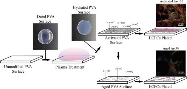
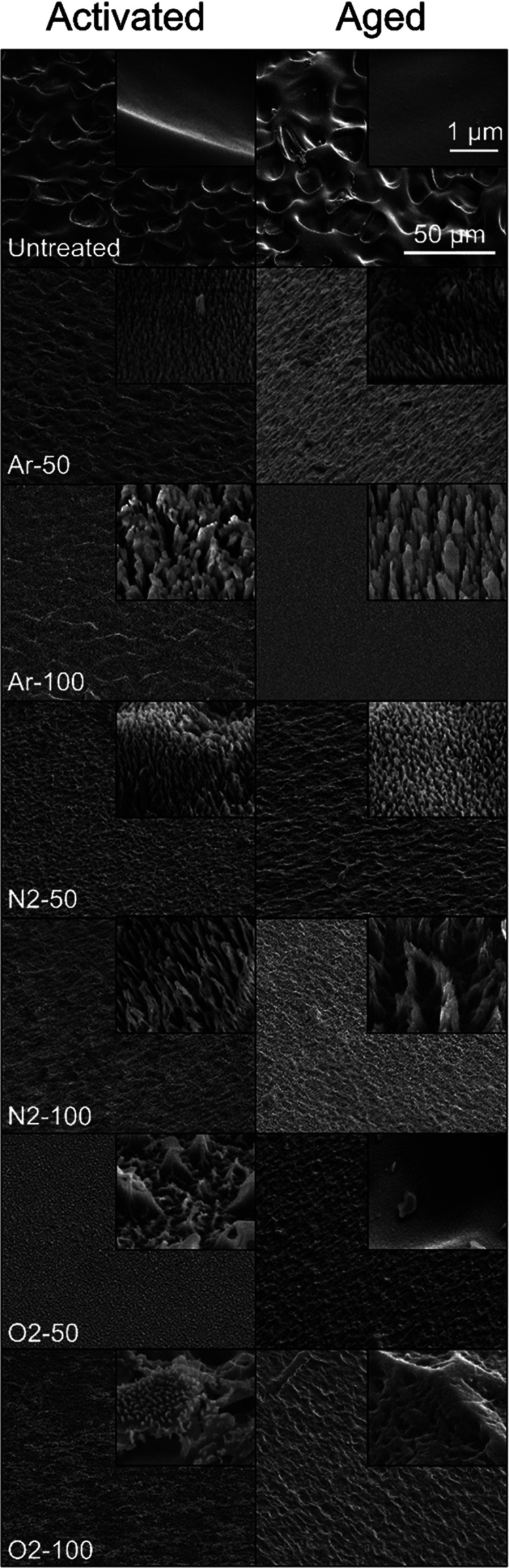
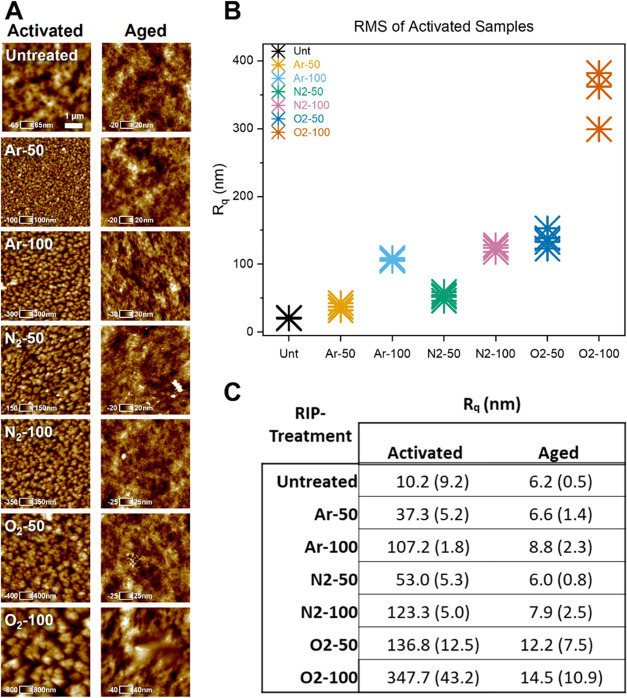
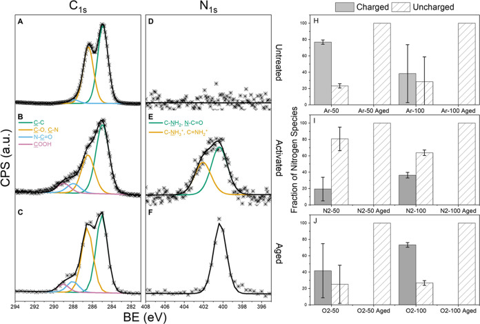
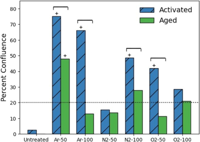
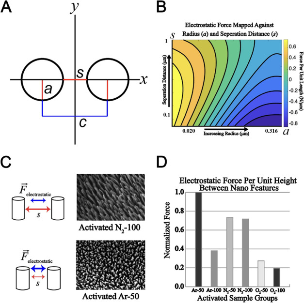
Synthetic small-diameter vascular grafts (<6 mm) are used in the treatment of cardiovascular diseases, including coronary artery disease, but fail much more readily than similar grafts made from autologous vascular tissue. A promising approach to improve the patency rates of synthetic vascular grafts is to promote the adhesion of endothelial cells to the luminal surface of the graft. In this study, we characterized the surface chemical and topographic changes imparted on poly(vinyl alcohol) (PVA), an emerging hydrogel vascular graft material, after exposure to various reactive ion plasma (RIP) surface treatments, how these changes dissipate after storage in a sealed environment at standard temperature and pressure, and the effect of these changes on the adhesion of endothelial colony-forming cells (ECFCs). We showed that RIP treatments including O2, N2, or Ar at two radiofrequency powers, 50 and 100 W, improved ECFC adhesion compared to untreated PVA and to different degrees for each RIP treatment, but that the topographic and chemical changes responsible for the increased cell affinity dissipate in samples treated and allowed to age for 230 days. We characterized the effect of aging on RIP-treated PVA using an assay to quantify ECFCs on RIP-treated PVA 48 h after seeding, atomic force microscopy to probe surface topography, scanning electron microscopy to visualize surface modifications, and X-ray photoelectron spectroscopy to investigate surface chemistry. Our results show that after treatment at higher RF powers, the surface exhibits increased roughness and greater levels of charged nitrogen species across all precursor gases and that these surface modifications are beneficial for the attachment of ECFCs. This study is important for our understanding of the stability of surface modifications used to promote the adhesion of vascular cells such as ECFCs.

Keywords: cardiovascular biomaterials; endothelialization; hydrophobic recovery; nanotopography; poly(vinyl alcohol); reactive ion plasma.


Conflict of interest statement

The authors declare no competing financial interest.

Figures

Figure 1
Figure 1
Schematic depicting the experimental procedure for quantifying ECFCs on activated or aged STMP-PVA surfaces. The hydrogel is cross-linked using sodium trimetaphosphate (STMP) and then subjected to reactive ion plasma (RIP) surface treatment in a dry state. This treatment introduces nanotopography and charged chemical species onto the surface, enhancing the endothelialization potential. Following the RIP treatment, the samples are hydrated for cell culture experiments. Over approximately 230 days of storage, the nanoscale surface roughness, charged chemical species, and endothelialization potential diminish. Representative fluorescence images of ECFCs cultured on activated Ar-100 and aged Ar-50 RIP-treated PVA are provided as insets. The images, captured at 20× magnification, are fluorescently labeled to indicate cell nuclei (blue), actin filaments (red), and VE-cadherin (green).
Figure 2
Figure 2
Scanning electron micrographs of untreated, activated, and aged PVA samples at 45° angle to the surface. The top row shows images of untreated PVA, the left-hand column shows images of activated PVA, and the right-hand column shows images of aged PVA. The order of RIP treatments from top to bottom is Untreated, Ar-50, Ar-100, N2-50, N2-100, O2-50, and O2-100. Larger images were collected at 1000× magnification with a 50 μm scale bar, and inset images were taken at 50 000× magnification with a 1 μm scale bar. RIP treatments impart nanohairs on the surface of the activated samples that are diminished in the aged samples. The qualitative character of the topographic surface modifications of activated and aged samples depends on the ion source gas and the power of the RIP treatment.
Figure 3
Figure 3
(A) Atomic force micrographs of activated and aged PVA samples for each RIP treatment and the untreated PVA. An in-plane scale bar of 1 μm is shown in the micrograph of untreated PVA, with orthogonal height values specific to each image. (B) Plot of the root-mean-square roughness (Rq) for each of the activated samples and the untreated polymer (n = 4). Activated samples at both RF powers were significantly different from the untreated samples with a one-sided paired t test (p < 0.05). (C) Rq values for activated and aged samples presented as the mean ± standard deviation. All activated samples returned to the approximate roughness of untreated PVA after aging for all RIP treatments, and were significantly different from activated samples according to ANOVA and Tukey’s post hoc test (p < 0.05).
Figure 4
Figure 4
(A–C), (D–F) (Left) High-resolution XPS scans of the C 1s and N 1s regions of PVA with N2-100 RIP treatment. The top row (A, D) represents unmodified PVA, the middle row (B, E) shows activated PVA after N2-100 RIP treatment, and the bottom row (C, F) shows aged PVA 230 days after N2-100 RIP treatment. The C 1s scans remained relatively unchanged between the activated and aged samples. In the N 1s region, a charged species of nitrogen was present in the activated sample but not in the aged sample. (H–J) (Right) The fraction of nitrogen species for each RIP treatment shown as either charged or uncharged nitrogen. The top row (H) shows the Ar-treated samples; the middle row (I) shows the N2-treated samples, and the bottom row (J) shows the O2-treated samples. The left side bar (solid gray) of each panel shows the activated samples, and the right bar (diagonal hatch) shows the aged samples. No charged nitrogen was detected in any aged samples. Measurements were conducted with a 100 μm diameter spot size, and three samples were measured for each treatment to calculate variance; the error bars indicate the standard deviation.
Figure 5
Figure 5
Bar graph of the percentage of endothelial colony-forming cell confluence on RIP-treated PVA samples 48 h after seeding. The blue bars with the diagonal hatch represent the activated samples, located left of center, while the green bars represent the aged samples, located right of center. The dotted line indicates the cell seeding density. Brackets represent significant differences between activated and aged samples for a single RIP treatment level using a one-sided paired t test. Bars marked with a “+” indicate a significant difference from untreated PVA according to ANOVA with Tukey’s post hoc (p < 0.05). After aging of the RIP-treated samples, the percentage of endothelial cell confluence decreased for all treatments compared to the activated samples.
Figure 6
Figure 6
Analysis of electrostatic forces within RIP-etched STMP-PVA nanohairs. (A) Uniform-charged cylinder model representing the nanohairs etched into the STMP-PVA. (B) Log-scale plot showcasing the forces generated by nanohairs, based on their radius (a) and separation distance (s). (C) SEM images captured normal to the substrate surface, used for model parameter calculation for each sample type. (D) Bar graph representing the estimated electrostatic forces across samples, normalized by the maximum predicted value (Activated Ar-50), to facilitate comparisons of the force for each RIP treatment type.

References

    1. Zeng W.; Li Y.; Wang Y.; Cao Y.. Tissue Engineering of Blood Vessels. In Encyclopedia of Tissue Engineering and Regenerative Medicine; Reis R. L., Ed.; Academic Press: Oxford, 2019; pp 413–424 10.1016/B978-0-12-801238-3.65848-8. - DOI
    1. Lüscher T. F. Vascular Biology of Coronary Bypass Grafts. Curr. Opin. Cardiol. 1991, 6 (6), 868–876. - PubMed
    1. Dohmen P. M.; Pruss A.; Koch C.; Borges A. C.; Konertz W. Six Years of Clinical Follow-up with Endothelial Cell–Seeded Small-Diameter Vascular Grafts during Coronary Bypass Surgery. J. Tissue Eng. 2013, 4, 2041731413504777 10.1177/2041731413504777. - DOI - PMC - PubMed
    1. Ma Q.; Shi X.; Tan X.; Wang R.; Xiong K.; Maitz M. F.; Cui Y.; Hu Z.; Tu Q.; Huang N.; Shen L.; Yang Z. Durable Endothelium-Mimicking Coating for Surface Bioengineering Cardiovascular Stents. Bioact. Mater. 2021, 6 (12), 4786–4800. 10.1016/j.bioactmat.2021.05.009. - DOI - PMC - PubMed
    1. Park K.-S.; Kang S. N.; Kim D. H.; Kim H.-B.; Im K. S.; Park W.; Hong Y. J.; Han D. K.; Joung Y. K. Late Endothelial Progenitor Cell-Capture Stents with CD146 Antibody and Nanostructure Reduce in-Stent Restenosis and Thrombosis. Acta Biomater. 2020, 111, 91–101. 10.1016/j.actbio.2020.05.011. - DOI - PubMed