. 2023 Dec 19;4(12):101326.
doi: 10.1016/j.xcrm.2023.101326.

PRMT blockade induces defective DNA replication stress response and synergizes with PARP inhibition

Affiliations

PRMT blockade induces defective DNA replication stress response and synergizes with PARP inhibition

Yang Li et al. Cell Rep Med. .

Abstract

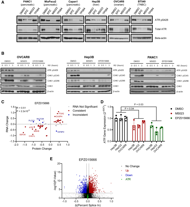
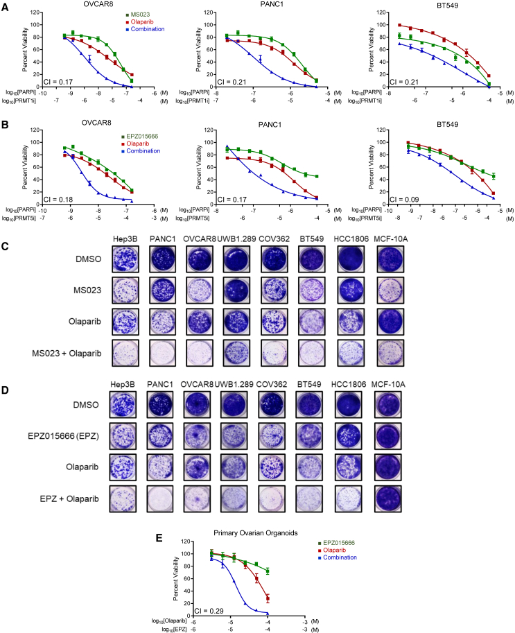
Multiple cancers exhibit aberrant protein arginine methylation by both type I arginine methyltransferases, predominately protein arginine methyltransferase 1 (PRMT1) and to a lesser extent PRMT4, and by type II PRMTs, predominately PRMT5. Here, we perform targeted proteomics following inhibition of PRMT1, PRMT4, and PRMT5 across 12 cancer cell lines. We find that inhibition of type I and II PRMTs suppresses phosphorylated and total ATR in cancer cells. Loss of ATR from PRMT inhibition results in defective DNA replication stress response activation, including from PARP inhibitors. Inhibition of type I and II PRMTs is synergistic with PARP inhibition regardless of homologous recombination function, but type I PRMT inhibition is more toxic to non-malignant cells. Finally, we demonstrate that the combination of PARP and PRMT5 inhibition improves survival in both BRCA-mutant and wild-type patient-derived xenografts without toxicity. Taken together, these results demonstrate that PRMT5 inhibition may be a well-tolerated approach to sensitize tumors to PARP inhibition.

Keywords: DNA replication stress; PARP inhibitors; PRMT inhibitors; arginine methylation; breast cancer; ovarian cancer.


Conflict of interest statement

Declaration of interests M.T.L. is a founder and limited partner in StemMed, Ltd., and a manager in StemMed Holdings, its general partner. He is a founder and equity stakeholder in Tvardi Therapeutics, Inc. Some PDXs are exclusively licensed to StemMed, Ltd., resulting in royalty income to M.T.L. L.E.D. is a compensated employee of StemMed, Ltd. Some PDXs are exclusively licensed to StemMed, Ltd., resulting in royalty income to L.E.D. A.K.S. discloses the following competing interests: consulting (Merck, GSK, ImmunoGen, Iylon, Kiyatec, Astra Zeneca, Onxeo), shareholder (BioPath), and patent (EGFL6 antibody).

Figures

None
Graphical abstract
Figure 1
Figure 1
Targeted proteomics reveal landscape of signaling changes following PRMT inhibition (A) Overview of experimental design. Twelve cell lines (Table S1) were treated with PRMT inhibitors targeting PRMT1 (MS023, 5 μM), PRMT4 (TP064, 10 μM), PRMT5 (EPZ015666, 5 μM), or DMSO vehicle control for 5 days prior to harvesting for targeted proteomics by RPPA in duplicate. (B) Correlation in PRMT-inhibitor (PRMTi)-induced changes in protein expression between MS023, TP064, and EPZ015666. Pearson correlation coefficient. (C) Overlap of significantly (false discovery rate [FDR] < 0.1) differentially expressed proteins between different inhibitors. Significance was determined using a generalized mixed linear model, taking each cell line as an independent biological replicate (N = 12). (D) All proteins significantly altered by at least one PRMTi as described in (C). Color indicates magnitude of protein change ranging from red (up-regulated) to white (no change) to blue (down-regulated). Dots with black circles indicate significant alterations (FDR < 0.1). Values are sorted by average magnitude of PRMTi-induced changes as determined by regression coefficient and divided based on if the average change is positive (left, top) or negative (right) or if it varied highly across inhibitors (left, bottom).
Figure 2
Figure 2
PRMT inhibition suppresses ATR expression, preventing activation of DNA replication stress response (A) Western blot to validate suppression of total ATR and ATR pS428 following 5 days of treatment with indicated PRMTis or DMSO vehicle control in indicated cell lines. (B) Western blot to assess ability of cells to activate CHK1 in response to exogenous replication stress. Cells were pre-treated for 5 days with indicated PRMTis or DMSO vehicle control and then exposed to 2 mM hydroxyurea (HU) for indicated times. (C) Scatterplot showing correlation between changes following PRMT5 inhibition at the protein level (RPPA) and transcriptional level (RNA sequencing [RNA-seq]). Each dot indicates a total protein significantly altered by PRMT5 inhibition. Genes with significantly altered expression following PRMTi are highlighted either in red (consistent with protein changes) or blue (inconsistent with protein changes). Inset R indicates Spearman correlation coefficient. RNA-seq was performed in duplicate per cell line, taking each cell line as an independent biological replicate (N = 4). (D) Gene expression changes as assessed by RT-qPCR following PRMT inhibition for 5 days in indicated cell lines. Gene expression was assessed as 2−ΔΔCt, where ΔCt was first assessed relative to internal 18s rRNA control and then normalized to DMSO-vehicle-treated samples. Mean ± SD, each dot represents a biological replicate. Statistics were performed taking each cell line as an independent observation (N = 3). Student’s t test. (E) Analysis of alternative splicing following treatment with EPZ015666, with significance taken as FDR <0.1 and |Δ(percent spliced in)| >0.05. Alternative splicing detected in ATR is highlighted by green dots but failed to reach significance. Four cell lines were analyzed in duplicate, and significance was determined by analyzing all 4 together, taking cell line as a batch effect. RNA-seq was performed in duplicate per cell line, taking each cell line as an independent biological replicate (N = 4).
Figure 3
Figure 3
PRMT inhibition potentiates PARPi-induced DNA damage (A) Immunocytochemistry in OVCAR8 cells for single-strand DNA (ssDNA) breaks in green as detected by native CldU, double-strand DNA breaks/general DNA damage in red as detected by γH2AX, cells synthesizing DNA in blue as detected by EdU, and DAPI counterstain in white. Superimposed white borders indicate segmented nuclei boundaries. Prior to foci staining, cells were treated with indicated PRMTis for 5 days followed by olaparib for 6 h. Scale bar, 10 μm. (B) Quantification of stains in (A). Only cells actively synthesizing DNA (EdU+) were used for quantification. Inset percentages indicate percentage of cells staining positive in each condition. Each dot in the volcano plot represents an individual cell, n > 1,000 cells per condition. Representative quantification from one replicate shown, n = 3 replicates per cell line. Shaded region indicates threshold for negative/positive staining. (C) Statistical analysis of data in (B) using a generalized linear mixed model with average values for each cell line (N = 3), taking cell line as a random effect. Error bars represent SE from regression model.
Figure 4
Figure 4
Blockade of PRMT activity synergizes with PARP inhibition (A) Cell lines were treated with indicated concentrations of the PRMT1i MS023, the PARPi olaparib, or combination thereof for 6 days before assessing cell viability relative to DMSO-vehicle-treated control. CI indicates combination index as assessed by the method of Chou and Talalay. See Figure S3A. (B) Cell lines were treated with indicated concentrations of the PRMT5i EPZ015666, the PARPi olaparib, or combination thereof for 6 days before assessing cell viability relative to DMSO-vehicle-treated control. CI indicates combination index as assessed by the method of Chou and Talalay. See Figure S3A. (C) Indicated cell lines were treated with the PRMT1i MS023, the PARPi olaparib, or combination thereof for 14 days before fixing and staining with crystal violet to visualize cell viability. Treatment concentrations and cell line annotations are given in Table S1. (D) Indicated cell lines were treated with the PRMT5i EPZ015666, the PARPi olaparib, or combination thereof for 14 days before fixing and staining with crystal violet to visualize cell viability. Treatment concentrations and cell line annotations are given in Table S1. (E) Primary patient-derived ovarian cancer organoids were treated with indicated concentrations of the PRMT5i EPZ015666, the PARPi olaparib, or combination thereof for 6 days before assessing cell viability relative to DMSO-vehicle-treated control. CI indicates combination index as assessed by the method of Chou and Talalay. Experiments were conducted in at least independent duplicate for every cell line.
Figure 5
Figure 5
Combination of the PRMT5i EPZ015666 and the PARPi olaparib is effective and well tolerated in vivo (A) The triple-negative breast cancer patient-derived xenograft BCM-7482 was allowed to reach ∼175 mm3 before treatment initiation with vehicle control (N = 8), 50 mg/kg olaparib 3× weekly (N = 8), 200 mg/kg EPZ015666 5× weekly (N = 7), or combination thereof (N = 7). Treatment was continued for 6 weeks or until mice reached maximum tumor volume. Log-rank test. (B) The ovarian patient-derived xenograft OV2428 was allowed to reach ∼175 mm3 before treatment initiation with vehicle control, 50 mg/kg olaparib 3× weekly, 200 mg/kg EPZ015666 5× weekly, or combination thereof. Treatment was continued for 6 weeks or until mice reached maximum tumor volume. Log-rank test. N = 5 per arm. (C) Bodyweights during treatment up until median survival time (25 days) of vehicle-control-treated mice, followed by bodyweights at endpoint for each treatment arm after axis break. Mean ± SD. (D) Hematocrit or red blood cell (RBC) count at either endpoint or time of treatment cessation in mice given indicated treatments. Each dot represents and individual mouse. Mean ± SD. (E) White blood cell (WBC) count at either endpoint or time of treatment cessation in mice given indicated treatments. Each dot represents and individual mouse. Mean ± SD. (F) Individual immune cell counts at either endpoint or time of treatment cessation in mice given indicated treatments. Each dot represents and individual mouse. Mean ± SD.

References

    1. Wang W.-H., Yuan T., Qian M.-J., Yan F.-J., Yang L., He Q.-J., Yang B., Lu J.-J., Zhu H. Post-translational modification of KRAS: potential targets for cancer therapy. Acta Pharmacol. Sin. 2021;42:1201–1211. - PMC - PubMed
    1. Fuentes-Antrás J., Alcaraz-Sanabria A.L., Morafraile E.C., Noblejas-López M.D.M., Galán-Moya E.M., Baliu-Pique M., López-Cade I., García-Barberán V., Pérez-Segura P., Manzano A., et al. Mapping of Genomic Vulnerabilities in the Post-Translational Ubiquitination, SUMOylation and Neddylation Machinery in Breast Cancer. Cancers. 2021;13 - PMC - PubMed
    1. Kumar M., Joshi G., Chatterjee J., Kumar R. Epidermal Growth Factor Receptor and its Trafficking Regulation by Acetylation: Implication in Resistance and Exploring the Newer Therapeutic Avenues in Cancer. Curr. Top. Med. Chem. 2020;20:1105–1123. - PubMed
    1. Shen Y., Gao G., Yu X., Kim H., Wang L., Xie L., Schwarz M., Chen X., Guccione E., Liu J., et al. Discovery of First-in-Class Protein Arginine Methyltransferase 5 (PRMT5) Degraders. J. Med. Chem. 2020;63:9977–9989. - PMC - PubMed
    1. Hwang J.W., Cho Y., Bae G.-U., Kim S.-N., Kim Y.K. Protein arginine methyltransferases: promising targets for cancer therapy. Exp. Mol. Med. 2021;53:788–808. - PMC - PubMed

Publication types

MeSH terms