. 2024 Feb;59(3):323-332.
doi: 10.1111/ejn.16232. Epub 2023 Dec 20.

Analysis of potassium ion diffusion from neurons to capillaries: Effects of astrocyte endfeet geometry

Affiliations

Analysis of potassium ion diffusion from neurons to capillaries: Effects of astrocyte endfeet geometry

Sara Djurich et al. Eur J Neurosci. 2024 Feb.

Abstract

Neurovascular coupling (NVC) refers to a local increase in cerebral blood flow in response to increased neuronal activity. Mechanisms of communication between neurons and blood vessels remain unclear. Astrocyte endfeet almost completely cover cerebral capillaries, suggesting that astrocytes play a role in NVC by releasing vasoactive substances near capillaries. An alternative hypothesis is that direct diffusion through the extracellular space of potassium ions (K+ ) released by neurons contributes to NVC. Here, the goal is to determine whether astrocyte endfeet present a barrier to K+ diffusion from neurons to capillaries. Two simplified 2D geometries of extracellular space, clefts between endfeet, and perivascular space are used: (i) a source 1 μm from a capillary; (ii) a neuron 15 μm from a capillary. K+ release is modelled as a step increase in [K+ ] at the outer boundary of the extracellular space. The time-dependent diffusion equation is solved numerically. In the first geometry, perivascular [K+ ] approaches its final value within 0.05 s. Decreasing endfeet cleft width or increasing perivascular space width slows the rise in [K+ ]. In the second geometry, the increase in perivascular [K+ ] occurs within 0.5 s and is insensitive to changes in cleft width or perivascular space width. Predicted levels of perivascular [K+ ] are sufficient to cause vasodilation, and the rise time is within the time for flow increase in NVC. These results suggest that direct diffusion of K+ through the extracellular space is a possible NVC signalling mechanism.

Keywords: astrocyte endfeet; diffusion; extracellular space; neurovascular coupling; potassium ions.


Figures

Figure 1.
Figure 1.
Schematic illustrations of extracellular pathways for diffusion of K+ and representation using model geometries. A. Diffusion of K+ from a local neuron (purple shape, arrows show release of K+). Yellow shapes represent other cells (neurons or neuroglia). The capillary is shown in cross-section in red with an external diameter of 5 μm. Astrocytes are shown in blue, with endfeet wrapped around the capillary. Gray represents the extracellular space. B. Idealized two-dimensional geometry representing local neuronal release of K+. Bold arrows show the boundary where K+ is released into the extracellular space, represented by concentration = 9 mM for t > 0. wc: width of cleft between astrocytic endfeet. wp: width of perivascular space. C. Diffusion of K+ from a neuron at a typical distance of 15 μm from vessel. Dotted curves show K+ diffusion pathways; otherwise as in A. D. Idealized two-dimensional geometry representing distant neuronal release of K+, otherwise as in B.
Figure 2.
Figure 2.
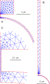
Meshes used for finite-element computations of K+ diffusion. A. Mesh used for local neuronal release geometry. B. Mesh used for distant neuronal release geometry. C. Enlarged view of cleft between astrocytic endfeet, showing fine mesh to resolve the cleft region.
Figure 3.
Figure 3.
Distribution of [K+] at four time points after K+ release for local neuronal release geometry, color-coded as indicated at right, with wc = 20 nm and wp = 100 nm. Initial concentration is 3 mM throughout the domain.
Figure 4.
Figure 4.
Variation in average perivascular [K+] over time following release of K+ in local neuronal release geometry. A. Fixed perivascular space width wp = 100 nm, cleft width wc varies from 6.67 to 100 nm. B. Fixed cleft width wc = 20 nm, perivascular space width wp varies from 20 to 500 nm.
Figure 5.
Figure 5.
Color-coded distribution of [K+] at four time points after K+ release for distant neuronal release geometry, color-coded as indicated at right, with wc = 20 nm and wp = 100 nm. Initial concentration is 3 mM throughout the domain.
Figure 6.
Figure 6.
Variation in average perivascular [K+] over time following release of K+ in distant neuronal release geometry. A. Fixed perivascular space width wp = 100 nm, cleft width wc varies from 6.67 to 100 nm. B. Fixed cleft width wc = 20 nm, perivascular space width wp varies from 20 to 500 nm.

References

    1. Ambrosi G, Virgintino D, Benagiano V, Maiorano E, Bertossi M, & Roncali L (1995). Glial cells and blood-brain barrier in the human cerebral cortex. Italian Journal of Anatomy and Embryology = Archivio Italiano Di Anatomia Ed Embriologia, 100 Suppl 1, 177–184. http://europepmc.org/abstract/MED/11322290 - PubMed
    1. Berne RM, Rubio R, & Curnish RR (1974). Release of Adenosine from Ischemic Brain. Circulation Research, 35, 262–271. 10.1161/01.RES.35.2.262 - DOI
    1. Bertossi M, Virgintino D, Maiorano E, Occhiogrosso M, & Roncali L (1997). Ultrastructural and Morphometric Investigation of Human Brain Capillaries in Normal and Peritumoral Tissues. Ultrastructural Pathology, 21, 41–49. 10.3109/01913129709023246 - DOI - PubMed
    1. Caesar K, Akgören N, Mathiesen C, & Lauritzen M (1999). Modification of activity-dependent increases in cerebellar blood flow by extracellular potassium in anaesthetized rats. The Journal of Physiology, 520, 281–292. 10.1111/j.1469-7793.1999.00281.x - DOI - PMC - PubMed
    1. Castejón OJ (1988). Ultrastructural alterations of human cortical capillary basement membrane in perifocal brain edema. Journal of Submicroscopic Cytology and Pathology, 20, 519–536. http://europepmc.org/abstract/MED/3179992 - PubMed

LinkOut - more resources