Review
. 2023 Dec 20;32(170):230138.
doi: 10.1183/16000617.0138-2023. Print 2023 Dec 31.

Clinical-radiological-pathological correlation in pulmonary arterial hypertension

Affiliations
Review

Clinical-radiological-pathological correlation in pulmonary arterial hypertension

Robin Condliffe et al. Eur Respir Rev. .

Abstract

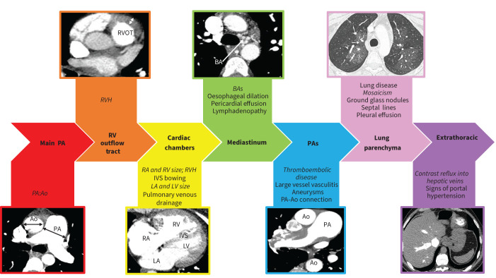
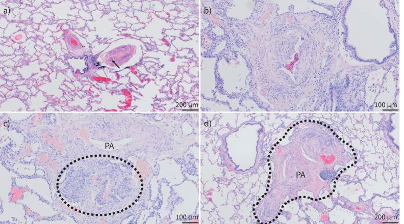
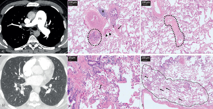
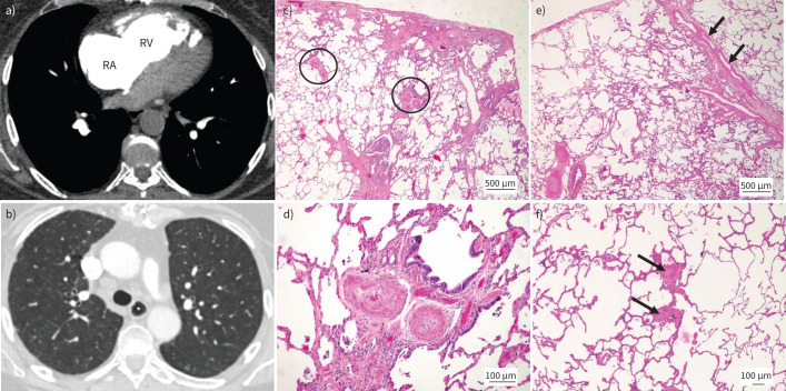
Pulmonary hypertension (PH) is defined by the presence of a mean pulmonary arterial pressure >20 mmHg. Current guidelines describe five groups of PH with shared pathophysiological and clinical features. In this paper, the first of a series covering all five PH classification groups, the clinical, radiological and pathological features of pulmonary arterial hypertension (PAH) will be reviewed. PAH may develop in the presence of associated medical conditions or a family history, following exposure to certain medications or drugs, or may be idiopathic in nature. Although all forms of PAH share common histopathological features, the presence of certain pulmonary arterial abnormalities, such as plexiform lesions, and extent of co-existing pulmonary venous involvement differs between the different subgroups. Radiological investigations are key to diagnosing the correct form of PH and a systematic approach to interpretation, especially of computed tomography, is essential.


Conflict of interest statement

Conflicts of interest: R. Condliffe, C. Durrington, A. Hameed, R.A. Lewis, R. Venkateswaran, D. Gopalan and P. Dorfmüller report no conflicts of interest related to this work.

Figures

FIGURE 1
FIGURE 1
Systematic approach to computed tomography pulmonary angiogram interpretation. Ao: aorta; BA: bronchial artery; IVS: interventricular septum; LA: left atrium; LV: left ventricle; PA: pulmonary artery; RA: right atrium; RV: right ventricle; RVH: right ventricular hypertrophy; RVOT: right ventricular outflow tract. Main features demonstrated in each image are in italics.
FIGURE 2
FIGURE 2
Pulmonary vascular histopathology in idiopathic and hereditary pulmonary arterial hypertension (PAH). Lungs from patients suffering from idiopathic and hereditary PAH; haematoxylin and eosin staining. a) Well-preserved lung parenchyma with slender alveolar septa and extended alveolar spaces; note the central bronchiole with two pulmonary arterial (PA) branches: the left one with medial and intimal concentric thickening, the right one displaying near-occlusive concentric intimal fibrosis (arrow indicates remaining lumen) and moderate lymphocytic infiltrate. b) PA with eccentric intimal fibrosis and cushion-like intimal thickening, suggesting an organised thrombotic event or in situ thrombosis. c) Plexiform lesion (circled) arising from the adventitia of a PA (top) and representing a connection between systemic vasa vasorum and pulmonary vasculature. d) Singular millimetric fibro-vascular lesion in a patient with hereditary PAH (bone morphogenetic protein type II receptor mutation); this vascular and fibrotic conglomerate comprising systemic and pulmonary vessels measures about 1.5 mm (circled area). a) and d) magnification ×40; b) and c) magnification ×100.
FIGURE 3
FIGURE 3
Case 1: idiopathic pulmonary arterial hypertension. a) Dilated main pulmonary artery (PA). b) Centrilobular ground glass changes in the absence of interlobular septal thickening or mediastinal lymphadenopathy. c–f) Lungs from the same patient. Haematoxylin and eosin staining; magnification: c) and e) ×100; d) ×200; and f) ×40. c) Classic plexiform lesion (circled), with adjacent dilation lesions (arrowheads), representing an open anastomosis between the pulmonary and the systemic (bronchial) vasculature; note the completely occluded small PA with concentric intimal fibrosis (arrow); also note the lymphocytic follicle between bronchiole and larger PA (asterisk). d) Small arteriole (about 50 µm in diameter, arrow) with important muscularisation and constriction of its lumen. e) Septal vein (circled) displaying prominent collagen-rich intimal fibrosis that appears focally constrictive. f) Peripheral lung with limiting visceral pleura (bottom); the arrows indicate multiple remodelled microvessels <70 µm in diameter, down to the pre-capillary level; in fact, the alveolar septa between the arterioles and venules appear focally thickened (circled area) this is probably due to the microvasculopathy; together with an increase in intra-alveolar macrophages that can be depicted as well, these histologic changes would be the morphological correlate for ground glass opacities in panel b.
FIGURE 4
FIGURE 4
Pulmonary arterial hypertension (PAH) in association with systemic sclerosis. a) Dilated right atrium (RA) and right ventricle (RV). b) Prominent oesophagus (*). c–f) Lungs from a different patient suffering from systemic sclerosis-associated PAH. Haematoxylin and eosin staining; magnification: c) and e) ×20; d) ×100; and f) ×40. c) Peripheral lung parenchyma with mild subpleural fibrosis (top). Note heavily remodelled muscular-type pulmonary arteries (PAs) (circled) in preserved areas, independent of fibrosis. d) Magnification of right circled PA. Medial muscular thickening and important near-occlusive concentric nonlaminar intimal fibrosis is present. e) Remodelled PAs (bottom left) and septal veins with substantial smooth muscle cell hyperplasia (arrows). f) Muscularisation of small pulmonary arterioles (arrows) and thickening of alveolar septa to the left of the arterioles; these changes are vaguely reminiscent of PAH group 1.6 (pulmonary veno-occlusive disease/pulmonary capillary haemangiomatosis).
FIGURE 5
FIGURE 5
Eisenmenger syndrome secondary to patent ductus arteriosus. a) Dilated main pulmonary artery (PA). b) Large patent ductus arteriosus (*). c) Right atrial (RA) and right ventricular (RV) dilatation with severe RV hypertrophy. d) Centrilobular ground glass change. e–f) Lungs from two other patients with congenital heart disease-associated pulmonary arterial hypertension. Haematoxylin and eosin staining; magnification ×40 and ×20, respectively. e) PAs of a patient with Eisenmenger syndrome show classic plexiform lesions of muscular-type arteries; note the blood-filled dilatation lesions (arrows) in direct vicinity to the plexiform core. f) Pulmonary septal vein with important smooth muscle cell hyperplasia in another patient with Eisenmenger syndrome due to an open ductus arteriosus; in this case, venous remodelling (arrows) and even pulmonary capillary haemangiomatosis-like capillary proliferation (thicker alveolar septa beneath the septal vein, circled area) were more prominent than arterial changes, demonstrating that different patients with Eisenmenger syndrome may be associated with different phenotypes of pulmonary vascular disease.

Comment in

References

    1. Humbert M, Kovacs G, Hoeper MM, et al. ESC/ERS guidelines for the diagnosis and treatment of pulmonary hypertension. Eur Respir J 2022; 43: 3618–3731. doi: 10.1093/eurheartj/ehac237 - DOI - PubMed
    1. European Respiratory Society . Pulmonary hypertension: clinical–radiological–pathological case series. Date last accessed: 8 July 2023. Date last updated: 31 March 2023. www.ersnet.org/events/pulmonary-hypertension-clinical-radiological-patho...
    1. Leber L, Beaudet A, Muller A. Epidemiology of pulmonary arterial hypertension and chronic thromboembolic pulmonary hypertension: identification of the most accurate estimates from a systematic literature review. Pulm Circ 2021; 11: 2045894020977300. doi: 10.1177/2045894020977300 - DOI - PMC - PubMed
    1. Rich S, Dantzker DR, Ayres SM, et al. Primary pulmonary hypertension. A national prospective study. Ann Intern Med 1987; 107: 216–223. doi: 10.7326/0003-4819-107-2-216 - DOI - PubMed
    1. Benza RL, Miller DP, Gomberg-Maitland M, et al. Predicting survival in pulmonary arterial hypertension: insights from the Registry to Evaluate Early and Long-Term Pulmonary Arterial Hypertension Disease Management (REVEAL). Circulation 2010; 122: 164–172. doi: 10.1161/CIRCULATIONAHA.109.898122 - DOI - PubMed

MeSH terms