. 2023 Dec 20;14(1):8465.
doi: 10.1038/s41467-023-43945-1.

Inflammasome activity is controlled by ZBTB16-dependent SUMOylation of ASC

Affiliations

Inflammasome activity is controlled by ZBTB16-dependent SUMOylation of ASC

Danfeng Dong et al. Nat Commun. .

Abstract

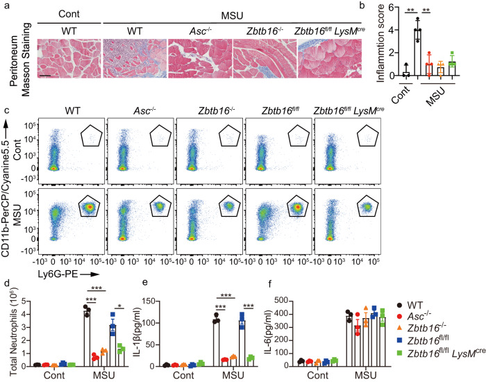
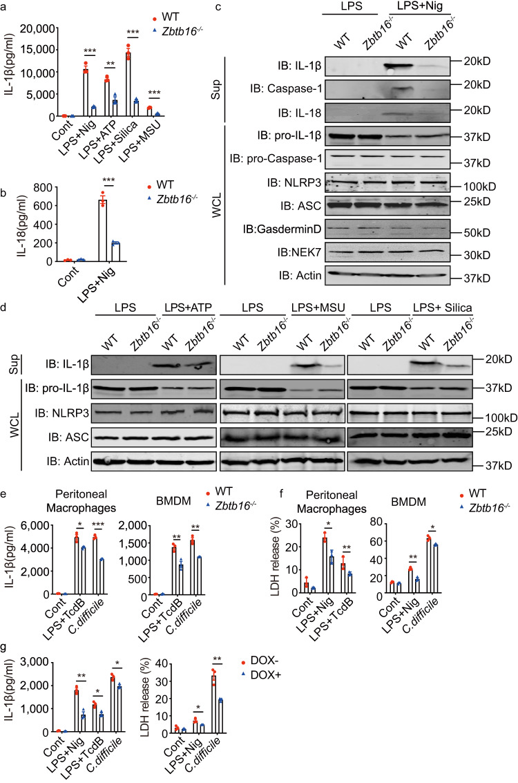
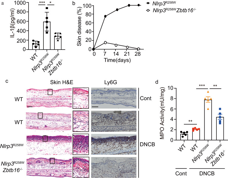
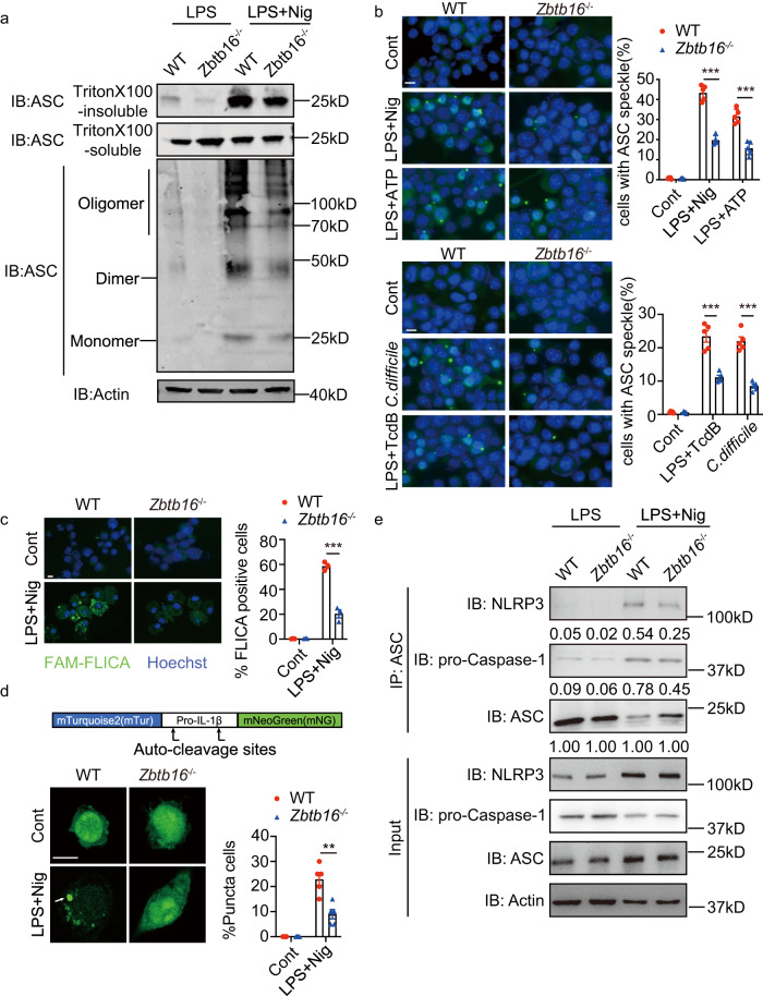
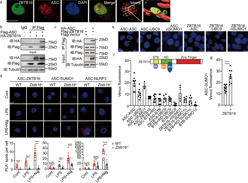
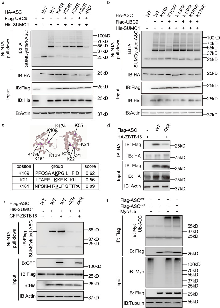
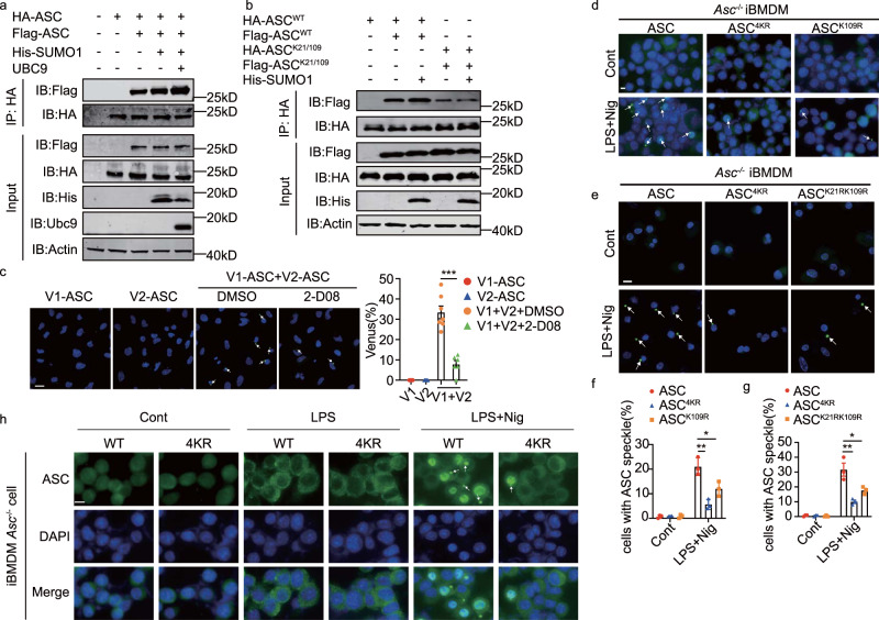
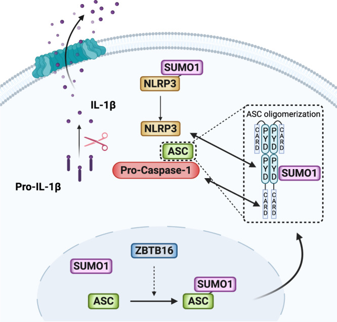
Inflammasome activity is important for the immune response and is instrumental in numerous clinical conditions. Here we identify a mechanism that modulates the central Caspase-1 and NLR (Nod-like receptor) adaptor protein ASC (apoptosis-associated speck-like protein containing a CARD). We show that the function of ASC in assembling the inflammasome is controlled by its modification with SUMO (small ubiquitin-like modifier) and identify that the nuclear ZBTB16 (zinc-finger and BTB domain-containing protein 16) promotes this SUMOylation. The physiological significance of this activity is demonstrated through the reduction of acute inflammatory pathogenesis caused by a constitutive hyperactive inflammasome by ablating ZBTB16 in a mouse model of Muckle-Wells syndrome. Together our findings identify an further mechanism by which ZBTB16-dependent control of ASC SUMOylation assembles the inflammasome to promote this pro-inflammatory response.


Conflict of interest statement

The authors declare no competing interests.

Figures

Fig. 1
Fig. 1. ZBTB16 promotes inflammasome-mediated pathogenesis.
WT, Asc-/-, Zbtb16fl/fl and Zbtb16 fl/flLysMCre mice were intraperitoneally injected with 3 mg monosodium urate (MSU) crystals and then assessed by (a) histological analysis of peritoneum from the indicated strains exposed to MSU processed by Masson staining to visualise pathogenesis (the scale bar = 50 μm) and (b) by an inflammation score, calculated as the sum of neo angiogenesis (0 when <3, 1 when 4–8, 2 when 9–12, 3 when >12 vessels) with polymorphonuclear cell (PMN) accumulation at the site of the injury (0 when <3, 1 when 4–8, 2 when 9–12, 3 when >12 activated PMNs are present) (n = 3 for control and n = 4 for MSU treated mice per group). After 6 h exposure to MSU peritoneal cells were collected from mice with 500 μl PBS lavage and assessed by counting the numbers of neutrophils (as CD11b+Ly6G+) by flow cytometry. The data are displayed as (c) representative dot plots and (d) a count of the total number of neutrophils in each condition. The levels of the inflammatory cytokines (e) IL-1β and (f) IL-6 in peritoneal fluids as measured by ELISA are shown as means ± SEM (n = 3 mice per group in each experiment). Two-tailed Student’s t-test were calculated for; WT vs Asc-/- p = 8.6 × 10−5 or 5.5 × 10−5, WT vs Zbtb16-/- p = 1.4 × 10−4 or 7.8 × 10−5 or Zbtb16fl/fl vs Zbtb16fl/flLysMCre p = 1.6 × 10−2 or 6.3 × 10−4 for the comparison of neutrophils or IL-1β, respectively (**p < 0.01 or ***p < 0.001). Source data are provided as a Source Data file.
Fig. 2
Fig. 2. ZBTB16 promotes inflammasome activation.
BMDMs from WT and Zbtb16-/- mice were either left untreated or primed with 1 μg/mL LPS for 4 h, followed by stimulation with 20 µM nigericin (Nig) or 5 mM ATP for 30 min, 120 μg/mL silica or 200 µg/mL MSU for 6 h, then the levels of the (a) IL-1β and (b) IL-18 cytokines were analysed in the supernatants by ELISA (n = 3, Data are presented as mean values ± SD) and (c, d) the cell lysates and supernatants were immunoblotted (IB) with the indicated antibodies. e IL-1β production by WT and Zbtb16-/- mouse peritoneal macrophages and BMDMs were primed with 1 μg/mL LPS for 2 h then stimulated with 200 ng/mL of TcdB toxin for 4 h or C. difficile 630 at MOI 100 for 6 h. f Cytotoxicity was assessed by LDH release in peritoneal macrophages and BMDMs treated with LPS then TcdB or 20 µM nigericin, or treated with C. difficile 630 for 30 min. g IL-1β production by THP-1 cells expressing ZBTB16 (DOX-) or ablated for ZBTB16 (DOX + , 1 µg/mL for 96 h) followed by treated with LPS and TcdB or nigericin or, alternatively, treated with C. difficile 630 (n = 3, data are presented as mean values ± SD for eg) was measured by ELISA in cell supernatants. Data are from at least three independent experiments. Statistical differences (*p < 0.05, **p < 0.01 or ***p < 0.001) were determined by a two-tailed Student’s t-test. Source data are provided as a Source Data file.
Fig. 3
Fig. 3. ZBTB16 controls a gain-of-function mutant inflammasome.
a The serum levels of IL-1β were examined from WT, Nlrp3R258W and Nlrp3R258WZbtb16- /- mice. b The percentage of Nlrp3R258W and Nlrp3R258WZbtb16-/- mice that developed skin inflammation within one month of birth is shown (n = 20). c, d 1-chloro-2,4-dinitrobenzene (DNCB) -induced skin inflammation in the indicated mice. c Representative H&E-stained sections of skin tissues showing the epidermal thickness and infiltration of neutrophils, as assessed by immunohistochemical staining with an anti-Ly6G antibody (scale bar = 75 μm) and by (d) measures of MPO activity as quantified by ELISA in skin extracts from untreated controls or 6 days after DNCB treatment. Error bars represent the mean ± SEM of technical replicates (n = 5 mice per group in each experiment). Statistical differences (**p < 0.01 or ***p < 0.001) were determined by a two-tailed Student’s t-test. Source data are provided as a Source Data file.
Fig. 4
Fig. 4. ZBTB16 promotes ASC oligomerization.
a BMDMs from WT and Zbtb16-/- mice were primed with 100 ng/mL LPS for 4 h and stimulated with nigericin followed by chemical cross-linking and centrifugation, then the pellets (Triton X 100-insoluble) and supernatants (Triton X 100-soluble) were probed by IB with an anti-ASC antibody. b Immunofluorescent detection of ASC specks in control untreated or BMDMs primed with LPS then stimulated with nigericin, ATP or TcdB, or treated with C. difficile alone and then stained with an anti-ASC antibody (green) and DAPI (blue) to visualised perinuclear ASC specks (scale bar = 10 μm) (n = 5). Representative micrographs are shown on the left and ASC specks in the cell cytosol are quantified on the right. c Caspase-1 activity in BMDMs primed with LPS and stimulated with nigericin is measured by FLICA assay (FAM-YVAD-FMK). Representative micrographs are shown on the left and quantification of FLICA-positive cells is graphed on the right as determined for 250 cells (scale bar = 10 μm) (n = 3). d Measures of the aggregation of a fluorescent-tagged pro-IL-1β as a cytosolic speck in BMDMs from WT and Zbtb16-/- mice transfected with the construct, then 24 h later stimulated with LPS and nigericin. The fluorescent signal in representative cells is shown on the left and the proportion of cells with GFP specks is quantified from 20 fluorescent positive cells in each field (scale bar = 10 μm). The percentage of cells with fluorescent puncta was scored in the graph on the right (n = 5). Data are presented as mean values ± SEM for (bd). e An assessment of the association of Asc with Nlrp3 and Caspase-1 by immunoprecipitation of lysates from treated BMDMs with an anti-ASC antibody then probing with the indicated fluorescently tagged antibodies. Quantification of each protein by their relative fluorescence in the IB is shown below the detected bands. All data are representative of at least three independent biological experiments. Statistical differences (**p < 0.01 or ***p < 0.001) were determined by a two-tailed Student’s t-test. Source data are provided as a Source Data file.
Fig. 5
Fig. 5. ZBTB16 promotes ASC SUMOylation.
a Detection of ASC SUMOylation by IB of lysates from HEK-293T cells co-transfected with HA-UBC9, Flag-ASC and His-SUMO1, His-SUMO2 or His-SUMO3, followed by precipitation with Ni-NTA resin to enrich SUMOylated proteins then probing with the indicated antibodies. Confirmation of the conjugation of SUMO1 to ASC by IB of lysates from HEK-293T cells co-transfected with tagged UBC9, ASC and SUMO1 and either (b) treated with the SUMOylation inhibitor 2-D08 (100 μM) enriched by HA beads or (c) by expression of the SUMO1 protease SENP1. SUMOylated proteins were enriched with Ni-NTA resin and then probed with the indicated antibodies. d Detection of ZBTB16-dependent promotion of ASC SUMOylation by IB of lysates from HEK-293T cells expressing CFP-ZBTB16, His-SUMO1 and Flag-ASC enrichment with Ni-NTA resin then probed with the indicated antibodies. e A confirmation that endogenous ASC SUMOylation is promoted by ZBTB16 in BMDM cells by IP of SUMOylated proteins from WT and Zbtb16-/- BMDM lysates with an anti-ASC antibody and then IB with an anti-SUMO1 antibody. f Measures of the effect of ZBTB16 on the colocalization of ASC and SUMO1 in WT and Zbtb16-/- BMDM cells untreated (Control) or primed with LPS for 4 h then treated with nigericin (LPS+Nig) to activate the Nlrp3 inflammasome. Representative micrographs showing single-plane confocal images of WT and Zbtb16- /- BMDMs cells with anti-ASC (green) and anti-SUMO1 (red) antibodies (scale bars = 10 µm). The extent of the colocalization of the ASC and SUMO1 signals is quantified in the graphs on the right with the IMARIS software (n = 9 for WT control, n = 6 for Zbtb16-/- control, n = 13 for WT stimulated and n = 16 for Zbtb16-/- stimulated, data are presented as mean values ± SD). Colocalization correlations and surface-to-surface localisation was determined in randomly selected cells over five fields. Each data point represented the mean value in the field. All data are representative of at least three independent experiments. Statistical differences (***p < 0.001) were determined by a two-tailed Student’s t-test. Source data are provided as a Source Data file.
Fig. 6
Fig. 6. ZBTB16 configures an ASC SUMOylation complex.
a Z-stack micrographs showing the nuclear location of ZBTB16 and ASC in BMDMs by immunofluorescence in cells transfecting with ZBTB16 and stained with DAPI (blue), mouse anti-ZBTB16 and rabbit anti-ASC primary antibodies followed by Alexa Fluor 488 anti-mouse (green) and Alexa Fluor 555 anti-rabbit (red) secondary antibodies. The corresponding reconstructed renderings by Imaris 9.5 (rightmost) for quantification of the contact area of ZBTB16 with ASC. Yellow indicated contact surface (scale bar = 10 µm). b, c Detection of an association between ZBTB16 and ASC by IB of lysates from HEK-293T cells co-transfected with tagged ASC and ZBTB16, immunoprecipitated (IP) with anti-Flag antibodies then probed with the indicated antibodies. d Representative fluorescent micrographs of WT and Zbtb16-/- BMDM cells stained by Proximity ligation assay (PLA) with antibodies for ASC and ZBTB16, SUMO1 or NLRP3. The cells were untreated (Cont) or primed with LPS for 4 h (LPS) and then stimulated with nigericin (LPS+Nig). The fluorescent signal is quantified in the graphs (below) as the mean ± s.e.m. Results are shown for three independent experiments with each data point representing 11−15 cells taken from 5 different views for each group. Cell nuclei are stained blue by DAPI (scale bar = 10 μm). e Fluorescent micrographs of HEK-293 cells expressing the indicated pairs of the following split-Venus tagged constructs: V1-ASC, V2-ASC, UBC9-V2, V1-SUMO1 (nSUMO1), SUMO1-V1 (SUMO1c), full-length V2-ZBTB16, ZBTB16 with the Zinc-finger domain removed (V2-BTB-RD) and just the BTB domain of ZBTB16 (V2-BTB). Cell nuclei are visualised by Hoechst staining (blue). As different exposure times were used to capture the separate images, the relative level of fluorescence is not representative (scale bars = 10 µm). f Total Venus fluorescence in HEK-293 cells co-transfected with the indicated split-Venus constructs and normalised against a negative control. The variance shows the standard error of the means from independent experiments (n = 8 for V1-ASC + V2-ASC, V1-UBC9 + V2-ASC, V1-ASC + V2-ZBTB16 / -RD-BTB / -BTB; n = 6 for V1-UBC9 + V2-ZBTB16 / -BTB-RD / -BTB, SUMO1-V1 + V2-ZBTB16 / -BTB-RD / -BTB; n = 4 for V1-SUMO1 + V2-ZBTB16; n = 3 for V1-SUMO1 + V2-ASC). The inset schematic depicts the predicted association of ASC, UBC9 and SUMO1 with the different protein domains of ZBTB16 by the bimolecular complementation. g A graph quantifying the relative levels of Venus fluorescence generated by an association between V1-SUMO1 and V2-ASC in HEK-293 cells co-transfected with constructs expressing UBC9 and either ZBTB16 or, as a control, the empty backbone vector (n = 9 per group, data are presented as mean ± SD). Statistical differences (***p < 0.001) were determined by a two-tailed Student’s t-test. Source data are provided as a Source Data file.
Fig. 7
Fig. 7. The lysine residue at position 21 and 109 on ASC is SUMOylated.
Measures of ASC SUMOylation in HEK-293T cells expressing UBC9, His-SUMO1 and the WT or mutant ASC constructs, with arginine replacement of the lysine residues (a) at positions 21 to 26 as separate (K21R, K22R, K24R and K26R) and combined (4KR) mutations or (b) positions 55 to 174 of HA-ASC. c A representation of the ASC structure (PDB: 2KN6) as a ribbon diagram showing the location of the lysine residues mutated in the study and (below) a table showing the results of a computational (SUMOplot) prediction of SUMOylated residues (underlined) on ASC that are calculated to have a high probability by scoring the match to a consensus sequence that binds UBC9 with substitution of counterpart residues. d Arginine replacement of the lysine residues between positions 21−26 on ASC is shown to affect its association with ZBTB16, as assessed in HEK-293T cells expressing HA-ZBTB16 and either the WT or the 4KR mutant Flag-ASC, followed by IP with an anti-HA antibody then IB with an anti-Flag antibody. e ZBTB16-dependent ASC SUMOylation is shown to depend on the lysine residues between positions 21−26, as shown by expressing UBC9, SUMO1 and ZBTB16 in HEK-293T cells with either the WT or mutant ASC (4KR), followed by precipitation of His-tagged SUMO1 with Ni-NTA resin, then IB for ASC with an anti-Flag antibody. The expression levels of all constructs are measured by IB with the indicated antibodies. f Measures of the ubiquitination of the lysine residues between positions 21−26 of ASC in HEK-293T cells expressing Myc-tagged ubiquitin (Myc-Ub) with either Flag-tagged WT or mutant (4KR) ASC, followed by IP with an anti-Flag antibody and IB with an anti-Myc antibody.
Fig. 8
Fig. 8. ASC SUMOylation and oligomerization are controlled by lysine residues at positions 21 and 109.
a Detection of the effect of SUMOylation for ASC multimerization in HEK-293T cells transfected with SUMO1, UBC9 and ASC differently tagged with Flag and HA, then IP with an anti-HA antibody followed by IB of SDS-PAGE gel separated immune-enriched proteins with an anti-Flag antibody. b Measures of the effect of arginine replacement of the lysine residues at positions 21 and 109 for ASC multimerization, as previously performed. c An assessment of the effect of SUMOylation on the oligomerization of ASC by measures of the reconstitution of a full-length fluorophore from each half of a split-Venus (V1 and V2) separately tagged ASC, either without (DMSO) or with a pharmacological inhibitor of SUMOylation (2-D08). Micrographs visualise ASC oligomers in the cytosol as Venus fluorescence (green) and DAPI-stained nuclei (blue) (scale bars = 10 μm) The relative proportion of cells with Venus fluorescence is quantified in the graphs (right) from randomly selected fields (n = 5 for V1 and V2, n = 7 for V1 + V2 with DMSO and n = 6 for V1 + V2 with 2-D08). Data are presented as mean values ± SD. dg Measures of the effect of the lysine residues between 21 and 26 and at position 109 on ASC oligomerization by expressing the indicated WT or arginine replacement mutant constructs (4KR, K109R and K21R + K109R) in immortalised Asc-/- BMDM treated with LPS and nigericin followed by detection of the formation of ASC specks with an anti-ASC antibody. Representative micrographs show the immunofluorescence pattern of the oligomerized ASC (green) in cells with DAPI-stained nuclei (blue) (scale bars = 10 μm). The effect of the different lysine residues on the formation of ASC specks is quantitated in at least 100 cells in 10 randomly selected fields. Data are presented as mean values ± SEM. h Micrographs showing the cellular distribution of exogenously expressed WT and the 4KR mutant ASC constructs expressed in immortalised Asc-/- BMDM at rest or upon priming with LPS followed by stimulation with nigericin (LPS+Nig) by immunofluorescent detection with an anti-ASC antibody (green). Cell nuclei were stained with DAPI (blue) (scale bars=10μm). Statistical differences (***p < 0.001) were determined by a two-tailed Student’s t-test (n = 3). Source data are provided as a Source Data file.
Fig. 9
Fig. 9. Inflammasome activity is promoted by ZBTB16-dependent SUMOylation of ASC.
A synopsis of the immune activity we report, in which inflammasome assembly progresses by a ZBTB16-SUMO1-ASC axis. ZBTB16 interacts with ASC in the nucleus to promote its modification with SUMO1. SUMOylated ASC then translocates to the cytosol where it may be deSUMOylated by SUMO proteases to re-enter the nucleus or, upon immune stimulus, assembles the inflammasome. We speculate that ASC SUMOylation alters its homologous and heterologous protein interactions and/or partitions ASC in the cytosol with other inflammasome constituents, some of which such as NLRP3 are also modified with SUMO1, to promote the inflammatory response.

References

    1. Latz E, Xiao TS, Stutz A. Activation and regulation of the inflammasomes. Nat. Rev. Immunol. 2013;13:397–411. doi: 10.1038/nri3452. - DOI - PMC - PubMed
    1. Broz P, Dixit VM. Inflammasomes: mechanism of assembly, regulation and signalling. Nat. Rev. Immunol. 2016;16:407–420. doi: 10.1038/nri.2016.58. - DOI - PubMed
    1. Guo H, Callaway JB, Ting JP. Inflammasomes: mechanism of action, role in disease, and therapeutics. Nat. Med. 2015;21:677–687. doi: 10.1038/nm.3893. - DOI - PMC - PubMed
    1. Christgen S, Place DE, Kanneganti TD. Toward targeting inflammasomes: insights into their regulation and activation. Cell Res. 2020;30:315–327. doi: 10.1038/s41422-020-0295-8. - DOI - PMC - PubMed
    1. Bryan NB, Dorfleutner A, Rojanasakul Y, Stehlik C. Activation of inflammasomes requires intracellular redistribution of the apoptotic speck-like protein containing a caspase recruitment domain. J. Immunol. 2009;182:3173–3182. doi: 10.4049/jimmunol.0802367. - DOI - PMC - PubMed

Publication types

MeSH terms