Review
. 2023 Dec 20;24(1):380.
doi: 10.1186/s12882-023-03438-1.

The functions of long non-coding RNA (lncRNA)-MALAT-1 in the pathogenesis of renal cell carcinoma

Affiliations
Review

The functions of long non-coding RNA (lncRNA)-MALAT-1 in the pathogenesis of renal cell carcinoma

Omid Anbiyaee et al. BMC Nephrol. .

Abstract

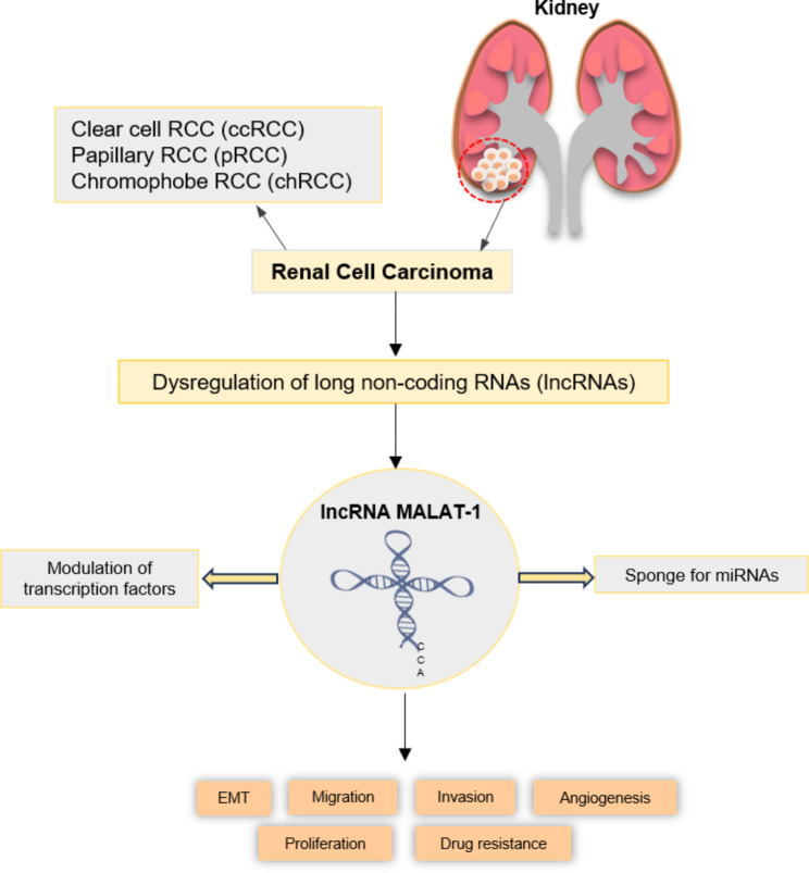
Renal cell carcinoma (RCC), a prevalent form of renal malignancy, is distinguished by its proclivity for robust tumor proliferation and metastatic dissemination. Long non-coding RNAs (lncRNAs) have emerged as pivotal modulators of gene expression, exerting substantial influence over diverse biological processes, encompassing the intricate landscape of cancer development. Metastasis-associated lung adenocarcinoma transcript 1 (MALAT-1), an exemplar among lncRNAs, has been discovered to assume functional responsibilities within the context of RCC. The conspicuous expression of MALAT-1 in RCC cells has been closely linked to the advancement of tumors and an unfavorable prognosis. Experimental evidence has demonstrated the pronounced ability of MALAT-1 to stimulate RCC cell proliferation, migration, and invasion, thereby underscoring its active participation in facilitating the metastatic cascade. Furthermore, MALAT-1 has been implicated in orchestrating angiogenesis, an indispensable process for tumor expansion and metastatic dissemination, through its regulatory influence on pro-angiogenic factor expression. MALAT-1 has also been linked to the evasion of immune surveillance in RCC, as it can regulate the expression of immune checkpoint molecules and modulate the tumor microenvironment. Hence, the potential utility of MALAT-1 as a diagnostic and prognostic biomarker in RCC emerges, warranting further investigation and validation of its clinical significance. This comprehensive review provides an overview of the diverse functional roles exhibited by MALAT-1 in RCC.

Keywords: Biomarker; MALAT-1; Renal cell carcinoma; lncRNAs.


Conflict of interest statement

The authors declare no competing interests.

Figures

Fig. 1
Fig. 1
The intricate biogenesis process of MALAT-1 and provides insights into its diverse functions in cancer cells. MALAT1, a long non-coding RNA, is transcribed by RNA polymerase II (Pol II). The RNA precursor of MALAT-1 undergoes specific cleavage mediated by RNase P, which occurs immediately downstream of the poly(A)-rich tract. This cleavage event results in the generation of the 3’ end of MALAT-1 and the 5’ end of a small RNA molecule resembling transfer RNA, known as MALAT-1-associated small cytoplasmic RNA (mascRNA). Subsequently, the 3’ end of mascRNA undergoes further cleavage by RNase Z, followed by the addition of a CCA sequence to the newly generated 3’ end. The mature 3’ end of MALAT-1 exhibits a distinct triple helix structure that sets it apart from the conventional poly(A) tail. This unique structure is attributed to the presence of an A-rich region encoded in the genome, along with two U-rich motifs. The triple helix structure contributes to the stability and functional properties of MALAT-1. MALAT-1 has been implicated in various biological processes and has garnered significant attention as a key player in the development and progression of cancer. Its involvement in gene regulation, cellular signaling, and nuclear organization highlights its multifaceted role in cellular physiology and disease pathogenesis
Fig. 2
Fig. 2
The intricate interplay between MALAT-1 and its downstream targets in renal cell carcinoma (RCC). The aberrant expression of MALAT-1 in RCC has been identified and linked to the advancement of tumors, invasion of surrounding tissues, and the formation of metastases. By functioning as a miRNA sponge and modulating the activity of transcription factors, MALAT-1 disrupts the normal gene expression profiles in RCC cells, thereby fostering the initiation and progression of the disease
Fig. 3
Fig. 3
MALAT-1 plays important roles in RCC development and progression

References

    1. Elballal MS, Sallam A-AM, Elesawy AE, Shahin RK, Midan HM, Elrebehy MA et al. miRNAs as potential game-changers in renal cell carcinoma: Future Clinical and Medicinal uses. Pathology-Research and Practice 2023:154439. - PubMed
    1. Boussios S, Devo P, Goodall IC, Sirlantzis K, Ghose A, Shinde SD, et al. Exosomes in the diagnosis and treatment of renal cell Cancer. Int J Mol Sci. 2023;24:14356. doi: 10.3390/ijms241814356. - DOI - PMC - PubMed
    1. Linehan WM, Ricketts CJ. The Cancer Genome Atlas of renal cell carcinoma: findings and clinical implications. Nat Reviews Urol. 2019;16:539–52. doi: 10.1038/s41585-019-0211-5. - DOI - PubMed
    1. di Meo NA, Lasorsa F, Rutigliano M, Milella M, Ferro M, Battaglia M, et al. The dark side of lipid metabolism in prostate and renal carcinoma: novel insights into molecular diagnostic and biomarker discovery. Expert Rev Mol Diagn. 2023;23:297–313. doi: 10.1080/14737159.2023.2195553. - DOI - PubMed
    1. Lucarelli G, Loizzo D, Franzin R, Battaglia S, Ferro M, Cantiello F, et al. Metabolomic insights into pathophysiological mechanisms and biomarker discovery in clear cell renal cell carcinoma. Expert Rev Mol Diagn. 2019;19:397–407. doi: 10.1080/14737159.2019.1607729. - DOI - PubMed

Substances