. 2023 Dec 20;11(1):206.
doi: 10.1186/s40478-023-01709-4.

Genetic ablation of Sarm1 attenuates expression and mislocalization of phosphorylated TDP-43 after mouse repetitive traumatic brain injury

Affiliations

Genetic ablation of Sarm1 attenuates expression and mislocalization of phosphorylated TDP-43 after mouse repetitive traumatic brain injury

Elif O Dogan et al. Acta Neuropathol Commun. .

Abstract

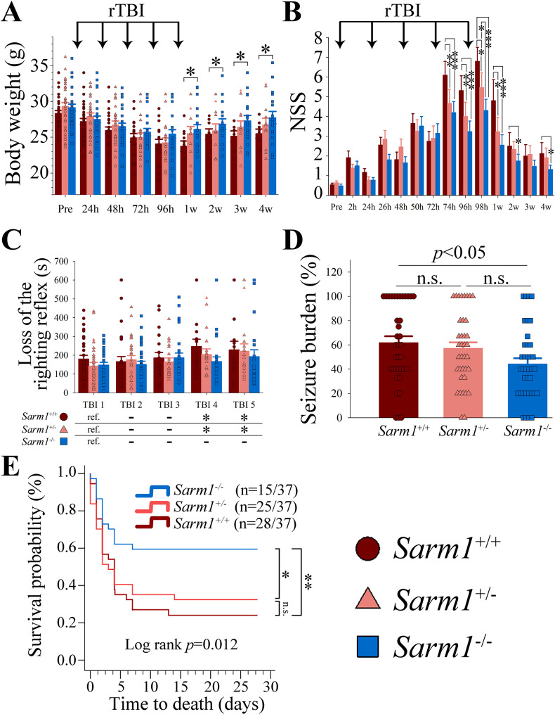
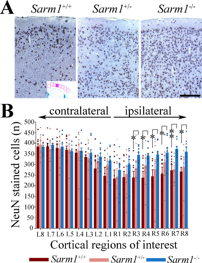
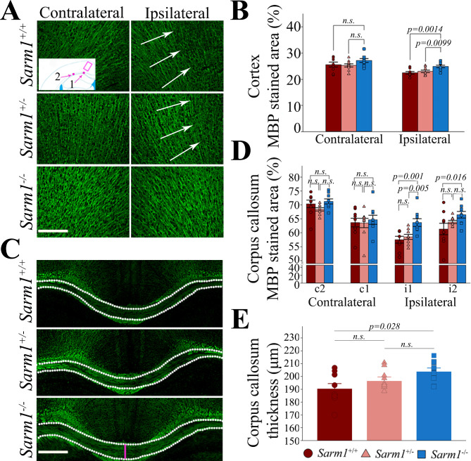
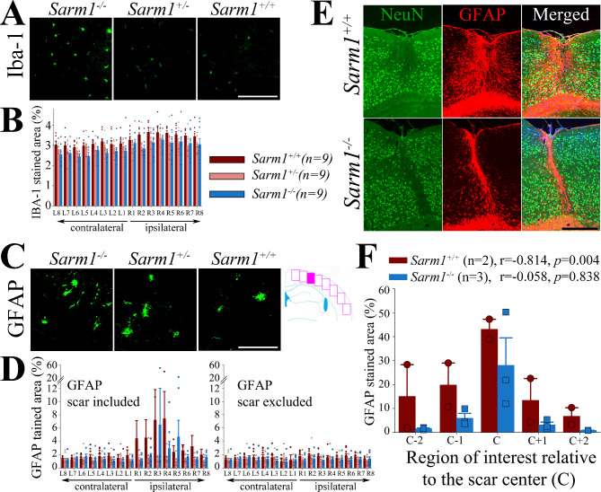
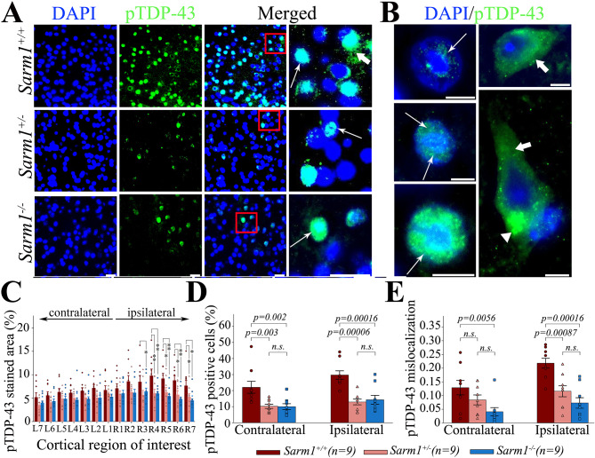
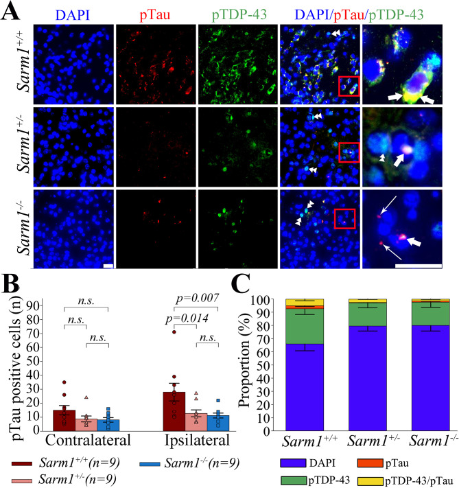
Traumatic brain injury (TBI), particularly when moderate-to-severe and repetitive, is a strong environmental risk factor for several progressive neurodegenerative disorders. Mislocalization and deposition of transactive response DNA binding protein 43 (TDP-43) has been reported in both TBI and TBI-associated neurodegenerative diseases. It has been hypothesized that axonal pathology, an early event after TBI, may promote TDP-43 dysregulation and serve as a trigger for neurodegenerative processes. We sought to determine whether blocking the prodegenerative Sarm1 (sterile alpha and TIR motif containing 1) axon death pathway attenuates TDP-43 pathology after TBI. We subjected 111 male Sarm1 wild type, hemizygous, and knockout mice to moderate-to-severe repetitive TBI (rTBI) using a previously established injury paradigm. We conducted serial neurological assessments followed by histological analyses (NeuN, MBP, Iba-1, GFAP, pTDP-43, and AT8) at 1 month after rTBI. Genetic ablation of the Sarm1 gene attenuated the expression and mislocalization of phosphorylated TDP-43 (pTDP-43) and accumulation of pTau. In addition, Sarm1 knockout mice had significantly improved cortical neuronal and axonal integrity, functional deficits, and improved overall survival after rTBI. In contrast, removal of one Sarm1 allele delayed, but did not prevent, neurological deficits and neuroaxonal loss. Nevertheless, Sarm1 haploinsufficient mice showed significantly less microgliosis, pTDP-43 pathology, and pTau accumulation when compared to wild type mice. These data indicate that the Sarm1-mediated prodegenerative pathway contributes to pathogenesis in rTBI including the pathological accumulation of pTDP-43. This suggests that anti-Sarm1 therapeutics are a viable approach for preserving neurological function after moderate-to-severe rTBI.

Keywords: Axon; Behavior; Brain injury; Glial scar; Haploinsufficiency; Interleukin; Neurodegeneration; SARM1; TDP-43; Tau.


Conflict of interest statement

The authors declare that they have no competing interests.

Figures

Fig. 1
Fig. 1
Genetic ablation of Sarm1, but not Sarm1 haploinsufficiency, mitigates rTBI-associated functional deficits and mortality. (A) While all rTBI groups lost weight after rTBI, Sarm1−/− mice recovered their weight faster than Sarm1+/+ and Sarm1+/−mice (p = 0.003 for group effects, p < 0.001 for time effects, p < 0.001 for group x time interaction). (B) While neurological deficits were significantly attenuated in both Sarm1−/− and mice Sarm1+/− up to 1 week post rTBI, only Sarm1−/− mice showed persistent protection up to the 4-week time point (p = 0.003 for group effects, p < 0.001 for time effects, p < 0.001 for group x time interaction). (C) Successive rTBI prolonged the time of the return of the righting reflex without difference between groups (group effect p = 0.219, time effect p = 0.016, group x time p = 0.600; *p < 0.05 versus TBI 1 [ref.]). (D) Sarm1−/− mice had a significantly lower seizure burden when compared to wild type mice (ANOVA on Ranks with post-hoc Tukey test). (E) Genetic ablation of Sarm1 significantly reduced rTBI-associated mortality. Numbers in parenthesis indicate the number of mice that died per the total number of mice in each group. Data in bar graphs are mean ± sem. *p < 0.05, **p < 0.01, ***p < 0.001
Fig. 2
Fig. 2
rTBI causes extensive neuronal loss at 1 month after injury that is attenuated by genetic ablation but not haploinsufficiency of Sarm1. (A) Loss of NeuN stained neurons in the cerebral cortex of Sarm1+/+ and Sarm1+/− mice after rTBI (images were taken from an area approximately corresponding to the filled square in the inset; rectangles indicate the approximate region of interest (ROI) used for quantitative analyses shown in panel [B]). (B) Although Sarm1+/− mice had numerically more neurons in the impacted hemisphere than Sarm1+/+ mice, this did not reach statistical significance. In contrast, genetic ablation of Sarm1 significantly suppressed neuronal loss (p = 0.028 for group effect, p < 0.001 for ROI, p = 0.010 for group x ROI interaction). Data in the bar graph are shown as mean ± sem. n = 9 per group. *p < 0.05. **p < 0.01. Scale bar = 50 μm
Fig. 3
Fig. 3
Genetic ablation of Sarm1 attenuates loss of myelin staining in the cerebral cortex and corpus callosum as well as mitigates corpus callosum atrophy at 1 month after rTBI. Representative myelin staining (MBP) from the (A) cerebral cortex and the (D) corpus callosum at 1 month after rTBI with corresponding quantified signal from (B) one region of interest (ROI) in the cerebral cortex (square in inset) and (C) two ROIs in the corpus callosum (dots in inset). Sarm1−/− mice had significantly greater MBP staining signal within the ipsilateral cortex when compared to Sarm1+/+ and Sarm1+/− (p < 0.001 for side effects, p = 0.032 for group effects, p = 0.513 for group x side interaction, arrows indicate region with attenuation of the MBP staining signal indicating axonal rarefaction). Similarly, genetic ablation of Sarm1 significantly attenuated myelin loss within the ipsilateral corpus callosum beneath the impact (ROI i1) and lateral to the impact (i2) (p = 0.023 for group effects, p < 0.001 for ROI effects, p = 0.079 for group x ROI interaction). There was no difference in myelin staining between groups in the corresponding contralateral ROIs (c1 and c2). (E) Sarm1−/− mice had less corpus callosum atrophy (measured in the mid-sagittal plane, pink line) when compared to Sarm1+/+ mice (One-way ANOVA with post hoc Holm-Šidák test). All data are mean ± sem; n = 9 per group. Scale bars = 200 μm
Fig. 4
Fig. 4
Genetic ablation of Sarm1 attenuates cortical microgliosis and astroglial scar formation. (A-B) At 1 month after rTBI, there were significantly more Iba-1-stained microglia in the injured versus non-injured cortex in all groups (p = 0.045 for group effects, p = 0.001 for ROI effects, p = 0.299 for group x ROI interaction), whereby this effect was attenuated in both Sarm1+/− (p = 0.028) and Sarm1−/− (p = 0.034) mice. (C-D) We observed focal astrogliosis in the injured cortex of Sarm1+/+ and Sarm1−/−, but not Sarm1+/−, mice. After exclusion of the glial scar from analysis, we found no difference in the GFAP-staining signal between hemispheres and groups (p = 0.145 for group effects, p = 0.071 for ROI effects, p = 0.605 for group x ROI interaction). (E) Representative micrographs showing the glial scar of Sarm1+/+ and Sarm1−/− mice with (F) corresponding quantification of the GFAP signal and correlation between GFAP and NeuN staining signal. Data in bar graphs are mean ± sem. n = 9 per group. Scale bars correspond to 100 μm in (A and C) and 500 μm in (E)
Fig. 5
Fig. 5
Sarm1 knockout and haploinsufficiency suppress TDP-43 pathology after rTBI. Representative micrographs showing (A) cortical pTDP-43 expression in Sarm1+/+, Sarm1−/−, and Sarm1+/− mice as well as (B) examples of neurons with different degrees of nuclear expression (long arrows), cytoplasmatic mislocalization (short arrows), and cytoplasmatic accumulation (arrowhead) of pTDP-43 in Sarm1+/+ mice. Compared to wild type Sarm1+/+ mice, Sarm1−/− and Sarm1+/− mice had significantly (C) attenuated pTDP-43 staining and (D) fewer pTDP-43 positive cells in the injured cerebral cortex at 1 month after rTBI. (E) Both Sarm1−/− and Sarm1+/− mice had significantly suppressed cytoplasmatic mislocalization of pTDP-43 in the injured cortex at 1 month. While there was also a reduction in pTDP-43 mislocalization within the non-impacted hemisphere, this effect was only significant in Sarm1−/− mice. Data are mean ± SEM; n = 9 per group. *p < 0.05, **p < 0.01. Scale bars correspond to 25 μm in (A) and 50 μm in (B)
Fig. 6
Fig. 6
Sarm1 knockout and haploinsufficiency suppress pTau expression after rTBI. (A) Representative micrographs showing the distribution of pTau versus pTDP-43 positive cells in the cerebral cortex at 1 month after rTBI. (B) Compared to wild type Sarm1+/+ mice, Sarm1−/− and Sarm1+/− mice had significantly fewer pTau positive cells in the injured cerebral cortex (long arrows indicate pTau stained cells, double arrowhead indicate pTDP-43 stained cells, and short arrow indicates pTDP-43/pTau double stained cells). (C) Shift towards fewer pTDP-43, pTau, and pTDP-43/pTau-double stained in Sarm1−/− and Sarm1+/− mice. While the proportion of pTDP-43 positive cells was similar for Sarm1−/− (17.0%) and Sarm1+/− (16.8%) groups, there were significantly fewer pTau and pTDP-43/pTau-double stained cells in Sarm1−/− (2.3%) versus Sarm1+/− (2.9%) (p < 0.05, χ2-test with post-hoc Bonferroni adjustment). Data are mean ± SEM; n = 9 per group. Scale bars = 50 μm

Similar articles

Cited by

References

    1. Alexandris AS, Koliatsos VE (2023) NAD(+), axonal maintenance, and neurological disease. 10.1089/ars.2023.0350. Antioxid Redox Signal - PMC - PubMed
    1. Alexandris AS, Lee Y, Lehar M, Alam Z, Samineni P, Tripathi SJ, et al. Traumatic axonopathy in spinal tracts after impact acceleration head injury: ultrastructural observations and evidence of SARM1-dependent axonal degeneration. Exp Neurol. 2023;359:114252. doi: 10.1016/j.expneurol.2022.114252. - DOI - PMC - PubMed
    1. Baughn MW, Melamed Z, Lopez-Erauskin J, Beccari MS, Ling K, Zuberi A, et al. Mechanism of STMN2 cryptic splice-polyadenylation and its correction for TDP-43 proteinopathies. Science. 2023;379:1140–1149. doi: 10.1126/science.abq5622. - DOI - PMC - PubMed
    1. Beers DR, Appel SH. Immune dysregulation in Amyotrophic Lateral Sclerosis: mechanisms and emerging therapies. Lancet Neurol. 2019;18:211–220. doi: 10.1016/S1474-4422(18)30394-6. - DOI - PubMed
    1. Bloom AJ, Mao X, Strickland A, Sasaki Y, Milbrandt J, DiAntonio A. Constitutively active SARM1 variants that induce neuropathy are enriched in ALS patients. Mol Neurodegeneration. 2022;17:1–15. doi: 10.1186/s13024-021-00511-x. - DOI - PMC - PubMed

Publication types