Review
. 2024 Feb 7;226(2):iyad200.
doi: 10.1093/genetics/iyad200.

Bone morphogenetic protein signaling: the pathway and its regulation

Affiliations
Review

Bone morphogenetic protein signaling: the pathway and its regulation

Takuya Akiyama et al. Genetics. .

Abstract

In the mid-1960s, bone morphogenetic proteins (BMPs) were first identified in the extracts of bone to have the remarkable ability to induce heterotopic bone. When the Drosophila gene decapentaplegic (dpp) was first identified to share sequence similarity with mammalian BMP2/BMP4 in the late-1980s, it became clear that secreted BMP ligands can mediate processes other than bone formation. Following this discovery, collaborative efforts between Drosophila geneticists and mammalian biochemists made use of the strengths of their respective model systems to identify BMP signaling components and delineate the pathway. The ability to conduct genetic modifier screens in Drosophila with relative ease was critical in identifying the intracellular signal transducers for BMP signaling and the related transforming growth factor-beta/activin signaling pathway. Such screens also revealed a host of genes that encode other core signaling components and regulators of the pathway. In this review, we provide a historical account of this exciting time of gene discovery and discuss how the field has advanced over the past 30 years. We have learned that while the core BMP pathway is quite simple, composed of 3 components (ligand, receptor, and signal transducer), behind the versatility of this pathway lies multiple layers of regulation that ensures precise tissue-specific signaling output. We provide a sampling of these discoveries and highlight many questions that remain to be answered to fully understand the complexity of BMP signaling.

Keywords: BMP signaling; DV patterning; Dpp; FlyBook; Gbb; NMJ; Sax; Tkv; morphogen gradient; wing patterning.


Conflict of interest statement

Conflicts of interest The author(s) declare no conflicts of interest.

Figures

Fig. 1.
Fig. 1.
Core BMP signaling components. a) BMPs are synthesized as large proproteins that form dimers, linked by a disulfide in the C-terminal domain. The bioactive ligand (as a homodimer or heterodimer) consists of the C-terminal ligand domain and the associated prodomain, depending on the site of proteolytic cleavage by a proprotein convertase, such as furin. All cleaved products can be secreted (Anderson and Wharton 2017). Distinct ligand forms of Gbb (Gbb15 and Gbb38) have been observed in vivo and shown to have different functions. b) BMP type I receptors Tkv and Sax form tetrameric complexes with type II receptors (Punt and Wit). The constitutively active type II receptor kinase phosphorylates serine residues in the GS domain of the type I receptor to activate its kinase. The intracellular R-Smad, Mad, is thus phosphorylated. Receptor complexes containing Tkv are competent to signal, while those containing only Sax fail to propagate a signal by phosphorylating Mad despite binding ligand. c) Smads Mad (R-Smad) and Medea (co-Smad) share a primary structure of MH1 and MH2 domains separated by a linker. Mad is phosphorylated on its C-terminal serines by the type I receptor kinase, while at sites within the linker by other kinases (see Fig. 4).
Fig. 2.
Fig. 2.
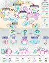
Regulation of BMP ligands. (Top) Posttranslational intracellular BMP regulation. Cell type–specific posttranslational modifications of inactive BMP proproteins occur in certain cellular organelles. After proper modifications, BMP producing cells secrete bioactive dimerized BMP ligands into the extracellular space. Several critical factors involved in this process are highlighted. The right column presents a simple diagram of BMP protein structures. (Bottom) Extracellular BMP regulation. Extracellular BMP interacting proteins control BMP ligand distribution in a context-dependent manner. GSC niche and NMJ exhibit a single-cell diameter BMP signaling range. On the other hand, BMP acts in intermediate and long distances during embryonic D/V patterning, PCV formation in the pupal wing, and longitudinal vein patterning in the developing wing disc.
Fig. 3.
Fig. 3.
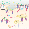
Receptor trafficking and degradation. BMP ligands interact with the ectodomain of type I and type II receptors in the extracellular space. Upon binding and activation of the type I receptor (star), the cytoplasmic Mad protein is phosphorylated prior to localizing to the nucleus where it regulates transcription (not shown). The ligand/receptor complex can be trafficked through different routes, first via clathrin-mediated or caveolae-mediated endocytosis. Nwk is thought to facilitate this process by binding to both Tkv and dynamin. Awd, Spict, and Scribb influence trafficking to the early endosome (EE) marked by Rab5 where active signaling has been observed. Asc1 facilitates trafficking to the recycling endosome (RE) marked by Rab11, from which receptors are thought to be delivered back to the cell surface. Alternatively, the receptor complex enters the late endosomal compartment (LE) destined for the lysosome (Lys) marked by Spin, trafficking mediated in part by Lgd and Shrub. Ube3 and Hiw, E3 ubiquitin ligases, have both been shown to target receptors to the proteosome. Ube3 preferentially enhances degradation of Tkv and not Sax or Wit. Nlg4 binds Tkv and prevents the action of S6KL and Smurf/Fu, both of whom have been shown to increase the degradation of Tkv.
Fig. 4.
Fig. 4.
Selected target sites for regulation of Mad activity and localization. All R-Smads and co-Smads share homology in 2 domains: MH1 and MH2. Mad has 2 N-terminal splice variants, conferring distinct N-terminal protein domains, shown here with the shorter Mad-PA N-terminus at the top left of MH1, and the longer Mad-PB N-terminus at the bottom left of MH1 (Sekelsky et al. 1995; Wiersdorff et al. 1996). The shorter isoform, Mad-PA, is commonly used for transgenic constructs, so the linker region amino acids are indicated by their location in Mad-PA. The linker region lies between MH1 and MH2 and has variable length across animal species, and only small stretches next to MH2 show sequence conservation between Mad and mammalian R-Smads. The MH1 domain contains the DNA binding site; the MH2 domain is involved in Smad–Smad association after phosphorylation. Stronger conservation is found within the R-Smads or within the co-Smads, which recognize distinct DNA binding sites. Inhibitory Smads share the MH2 domain but are divergent in the MH1 region (Hariharan and Pillai 2008). Known sites for the regulation of Mad activity mentioned herein are indicated as follows: Nemo-like kinase phosphorylation sites, S25 in Mad-PA or S95 in Mad-PB, are depicted in the N-terminal region. A proline-/serine-rich 34 amino acid sequence from the linker region is detailed. This short sequence contains 4 sites for proline-directed S/T kinase phosphorylation at 202, 212, 220 and 226. Among these, only phosphorylation at S212 has been studied in detail, but the proximity of additional sites raises the possibility that secondary sites could be targeted alternatively. Several of these sites can direct the kinase GSK3 (Sgg or Zw3 in Drosophila) to phosphorylate a nearby, more N-terminal serine. Linker phosphorylation is thought to recruit the binding of Smurf ubiquitylase to the nearby PPAY motif (underlined). The short C-terminal motif for activated BMP type I receptor phosphorylation is indicated as SSvS.
Fig. 5.
Fig. 5.
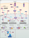
Regulation of Mad. Mad activity is regulated at the level of phosphorylation, nuclear-cytoplasmic shuttling, in association with transcriptional cofactors, as well as the mediator complex. a) Ligand–receptor interaction results in the phosphorylation of C-terminal serines. Two pCterMad associate with Medea, translocating to the nucleus to either activate or repress transcription. pCterMad is dephosphorylated by at least 3 phosphatases, PDP, MTMR4, and Dullard, which are each localized to different cellular compartments, nuclear, cytoplasmic, and associated with the nuclear envelope, respectively. Medea cycles in and out of the nucleus but is more likely to be retained when complexed with pCterMad. Mad is also phosphorylated by other kinases in its linker domain (Sgg and others not shown). Linker phosphorylation recruits Smurf, which facilitates ubiqutination and subsequent degradation of Mad. Linker phosphorylation also mediates association with Yki leading to different transcriptional outcomes. b) Sveral different BMP response elements have been identified, each mediating a different transcriptional response [BMP-AE, BMP silencer element (BMP-SE)]. c) Cdk8 is a component of the mediator kinase module and able to phosphorylate Smad1 and Mad linker domains. Mad and Smad1 are thought to integrate with Cdk8 and the mediator complex to influence transcription.

References

    1. Aberle H, Haghighi AP, Fetter RD, McCabe BD, Magalhães TR, Goodman CS. 2002. Wishful thinking encodes a BMP type II receptor that regulates synaptic growth in Drosophila. Neuron. 33(4):545–558. doi:10.1016/S0896-6273(02)00589-5. - DOI - PubMed
    1. Affolter M, Basler K. 2007. The Decapentaplegic morphogen gradient: from pattern formation to growth regulation. Nat Rev Genet. 8(9):663–674. doi:10.1038/nrg2166. - DOI - PubMed
    1. Affolter M, Marty T, Vigano MA, Jazwinska A. 2001. Nuclear interpretation of dpp signaling in Drosophila. EMBO J. 20(13):3298–3305. doi:10.1093/emboj/20.13.3298. - DOI - PMC - PubMed
    1. Affolter M, Nellen D, Nussbaumer U, Basler K. 1994. Multiple requirements for the receptor serine/threonine kinase thick veins reveal novel functions of TGF beta homologs during Drosophila embryogenesis. Development. 120(11):3105–3117. doi:10.1242/dev.120.11.3105. - DOI - PubMed
    1. Akiyama T, Gibson MC. 2015. Decapentaplegic and growth control in the developing Drosophila wing. Nature. 527(7578):375–378. doi:10.1038/nature15730. - DOI - PubMed

Publication types

MeSH terms

Substances

LinkOut - more resources