. 2023 Dec 21:29:e942137.
doi: 10.12659/MSM.942137.

Evaluation of Safety and Efficacy of Preoperative Coronal MRI-Guided Minimally Invasive Surgery for Cervical Spondylotic Radiculopathy

Affiliations

Evaluation of Safety and Efficacy of Preoperative Coronal MRI-Guided Minimally Invasive Surgery for Cervical Spondylotic Radiculopathy

Yuan Liu et al. Med Sci Monit. .

Abstract

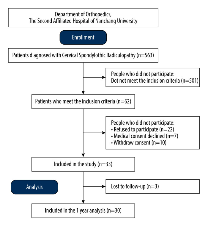
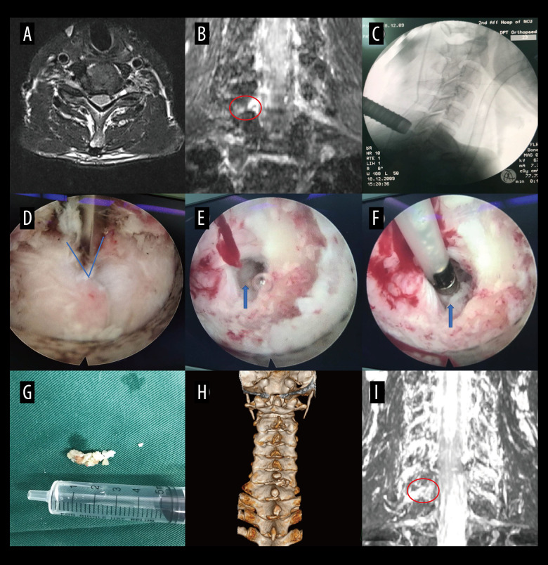
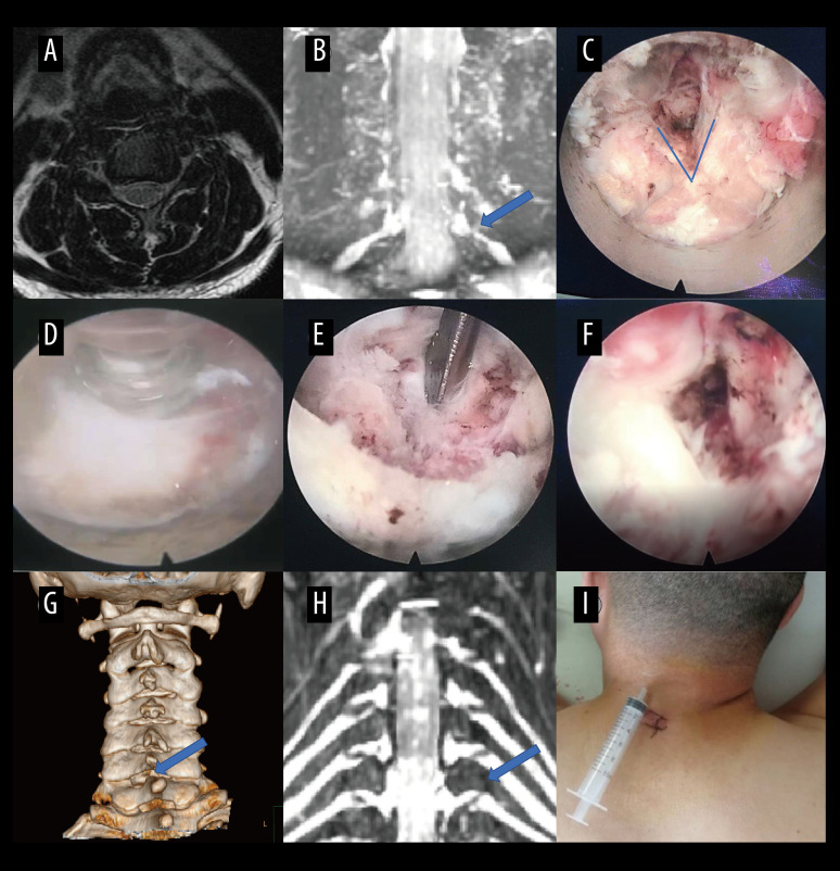
BACKGROUND Key-hole surgery is a minimally invasive technique that has shown promise in various surgical procedures. This study aimed to assess the clinical effectiveness of preoperative coronal MRI-assisted key-hole surgery for the treatment of patients with cervical spondylotic radiculopathy (CSR). MATERIAL AND METHODS A total of 30 patients diagnosed with CSR and undergoing key-hole surgery with CMRI assistance were included in the study. Various parameters, including surgical segments, incision length, disease duration, operative time, intraoperative fluoroscopy times, intraoperative blood loss, complications, and length of hospitalization, were recorded. Precise measurements of Cobb angles and intervertebral space height were taken before and after the surgical procedure. Surgical outcomes were evaluated using modified Macnab criteria, visual analogue scale (VAS), Japanese Orthopaedic Association Scores (JOA), and neck disability index (NDI). RESULTS The average duration of disease was 6.47±3.29 months, with an average incision length of 1.94±0.15 cm and operative time of 57.83±4.34 minutes. The average intraoperative blood loss was 33.70±9.28 ml, with an average of 3.50±0.73 intraoperative fluoroscopies. The average duration of hospitalization was 4.10±1.27 days. Preoperative and postoperative measurements showed no statistically significant difference in C2-C7 Cobb angles and intervertebral space height. However, there were significant improvements in postoperative VAS, NDI, and JOA scores compared to preoperative scores. The surgical effectiveness rate was 100%, with a high rate of good and excellent outcomes. CONCLUSIONS The findings of this study suggest that preoperative CMRI-assisted key-hole surgery for single-segment CSR is a safe and effective treatment option with low complication rates. The clinical benefits include high security and good outcomes. Further research and larger studies are warranted to validate these findings.


Conflict of interest statement

Conflict of interest: None declared

Figures

Figure 1
Figure 1
Flow diagram of enrollment, follow-up, and analysis.
Figure 2
Figure 2
Case 1: example of a typical case. (A) Coronal magnetic resonance image of 3-dimensional fast-field echo with water-selective excitation (CMRI) showed disc herniation at the left C7/T1 level, with nerve root filling defects (blue arrow). (B) Never root decompression performed, with good dural pulsation. (C) Postoperative CMRI showed good decompression of the left C8 nerve root (blue arrow). (D) Postoperative computed tomography image with appropriate windowing (blue arrow). Vue PACS, v12.2.6, Philips.
Figure 3
Figure 3
Case 2: example of a typical case. (A) Coronal magnetic resonance image of 3-dimensional fast-field echo with water-selective excitation (CMRI) showed disc herniation at the left C6/C7 level, with nerve root filling defects (blue arrow). (B) Surgical specimen of the removed intervertebral disc. (C) Postoperative CMRI showed good decompression of the right C7 nerve root (blue arrow). (D) Postoperative computed tomography image with appropriate windowing (blue arrow). Vue PACS, v12.2.6, Philips.
Figure 4
Figure 4
Case 3: example of a typical case. (A) Coronal magnetic resonance image of 3-dimensional fast-field echo with water-selective excitation (CMRI) showed foraminal stenosis at the left C6/C7 level (blue arrow). (B) Never root decompression performed. (C) Postoperative CMRI showed good decompression of the left C7 nerve root (blue arrow). (D) Postoperative computed tomography image with appropriate windowing (blue arrow). Vue PACS, v12.2.6, Philips.
Figure 5
Figure 5
A typical case: case 1. (A-D) Cervical spine magnetic resonance imaging (MRI) showed no significant cervical spinal canal stenosis in segments C4/C5, C5/C6, C6/C7, and C7/T1. (E) Coronal MRI of 3-dimensional fast-field echo with water-selective excitation (CMRI) showed disc herniation at the left C7/T1 level, with nerve root filling defects (blue arrow). (F) Preoperative CT imaging. (G) After pre-operative localization by CMRI imaging, insertion of a working cannula. (H) Exposure of the V-point (arrow). (I) Using a high-speed drilling to remove the vertebral plate and articular process around the V-point. (J) Never root decompression performed, with good dural pulsation. (K) Postoperative computed tomography image with appropriate windowing (blue arrow). (L) Postoperative CMRI showed good decompression of the left C8 nerve root (blue arrow). Vue PACS, v12.2.6, Philips.
Figure 6
Figure 6
A typical case: case 2. (A) Cervical spine magnetic resonance imaging (MRI) showed no apparent cervical spinal canal stenosis in segments C6/C7. (B) Coronal MRI of 3-dimensional fast-field echo with water-selective excitation (CMRI) showed disc herniation at the left C6/C7 level, with nerve root filling defects (red circle). (C) After pre-operative localization by CMRI imaging, insertion of a working cannula. (D) Exposure of the V-point (blue arrow). (E) Exposure of the protruding intervertebral disc (blue arrow). (F) Surgical specimen of the removed intervertebral disc. (G) Degenerated disc tissue for comparison. (H) Postoperative computed tomography image with appropriate windowing. (I) Postoperative CMRI showed good decompression of the right C7 nerve root (red circle). Vue PACS, v12.2.6, Philips.
Figure 7
Figure 7
A typical case: case 3. (A) Cervical spine magnetic resonance imaging (MRI) showed no apparent disc herniation in segments C6/C7. (B) Coronal MRI of 3-dimensional fast-field echo with water-selective excitation (CMRI) showed foraminal stenosis at the left C6/C7 level (blue arrow). (C) Exposure of the V-point. (D, E) Pictures during endoscopic surgery. (F) Never root decompression performed. (G) Postoperative computed tomography image with appropriate windowing (blue arrow). (H) Postoperative CMRI showed good decompression of the left C7 nerve root (blue arrow). (I) Postoperative small incision. Vue PACS, v12.2.6, Philips.

Similar articles

References

    1. Hu J, Chen F, Qiu G, et al. Jingshu Keli for treating cervical spondylotic radiculopathy: The first multicenter, randomized, controlled clinical trial. J Orthop Translat. 2021;27:44–56. - PMC - PubMed
    1. Luyao H, Xiaoxiao Y, Tianxiao F, et al. Management of cervical spondylotic radiculopathy: A systematic review. Global Spine J. 2022;12(8):1912–24. - PMC - PubMed
    1. Persson LC, Carlsson JY, Anderberg L. Headache in patients with cervical radiculopathy: A prospective study with selective nerve root blocks in 275 patients. Eur Spine J. 2007;16(7):953–59. - PMC - PubMed
    1. Benditz A, Brunner M, Zeman F, et al. Effectiveness of a multimodal pain management concept for patients with cervical radiculopathy with focus on cervical epidural injections. Sci Rep. 2017;7(1):7866. - PMC - PubMed
    1. Mazzucchi E, La Rocca G, Perna A, et al. Single-level anterior cervical discectomy and interbody fusion: A comparison between porous tantalum and polyetheretherketone cages. J Pers Med. 2022;12(6):986. - PMC - PubMed

MeSH terms