Review
. 2024 Apr;109(4):484-501.
doi: 10.1113/EP091586. Epub 2023 Dec 20.

Insights into pathophysiology and therapeutic strategies for heat stroke: Lessons from a baboon model

Affiliations
Review

Insights into pathophysiology and therapeutic strategies for heat stroke: Lessons from a baboon model

Mashan Abdullah et al. Exp Physiol. 2024 Apr.

Abstract

Heat stroke is a perilous condition marked by severe hyperthermia and extensive multiorgan dysfunction, posing a considerable risk of mortality if not promptly identified and treated. Furthermore, the complex biological mechanisms underlying heat stroke-induced tissue and cell damage across organ systems remain incompletely understood. This knowledge gap has hindered the advancement of effective preventive and therapeutic strategies against this condition. In this narrative review, we synthesize key insights gained over a decade using a translational baboon model of heat stroke. By replicating heat stroke pathology in a non-human primate species that closely resembles humans, we have unveiled novel insights into the pathways of organ injury and cell death elicited by this condition. Here, we contextualize and integrate the lessons learned concerning heat stroke pathophysiology and recovery, areas that are inherently challenging to investigate directly in human subjects. We suggest novel research directions to advance the understanding of the complex mechanisms underlying cell death and organ injury. This may lead to precise therapeutic strategies that benefit individuals suffering from this debilitating condition.

Keywords: cell death; coagulation abnormalities; heat stress response; hyperthermia; inflammatory response; organ injury.


Conflict of interest statement

The authors declare that they have no competing interests.

Figures

FIGURE 1
FIGURE 1
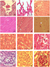
Microvascular alterations across organs in heat stroke. (a–c) Jejunum. (a) Normal intestinal villi in a control animal. (b) Moderate heat stroke shows limited villous capillary distension (arrow). (c) Severe heat stroke exhibits pronounced jejunal villous capillary swelling, red blood cell stasis (arrows), and amplified inflammatory cell presence with loss of surface epithelium. (d–f) Liver. (d) Control animal liver is unremarkable. (e) Moderate heat stroke shows sporadic platelets adhered to sinusoidal endothelium (arrowhead). (f) Extensive pan‐lobular sinusoidal hepatic congestion in severe heat stroke, spanning portal triads to central veins (CV), with amassing of leukocytes in sinusoids (arrows). (g–i) Lung. (g) Control animal lung is unremarkable. (h) Mild to moderate capillary congestion and leucocytes infiltration in moderate heat stroke (arrows). (i) Pronounced alveolar capillary distension with red blood cell engorgement (arrows), increased inflammatory cells, and partial arteriolar occlusion by thrombus (arrowhead) in severe heat stroke. (j–l) Heart. (j) Normal myocardial capillary in control. (k, l) Myocardial capillary engorgement (arrows) in both moderate (k) and severe (l) heat stroke. (m–o) Kidney. (m) Normal peritubular and glomerular capillaries in control. (n, o) Pronounced amplification and red blood cell accumulation of peritubular capillaries in moderate (arrows (n)) and severe (arrows (o)) heat stroke. Glomerular capillaries are engorged in severe heat stroke with expanded mesangial cellularity in both moderate (arrows (n)) and severe (arrows (o)) heat stroke. (p–r) Spleen. (p) Normal spleen architecture in control. (q) Moderate red pulp engorgement in moderate heat stroke (arrow). (r) Extensive red pulp congestion in severe heat stroke (straight arrows) with extension into the white pulp (curved arrow). (s–u) Adrenal. (s) Normal adrenal gland in control. (t, u) Pronounced sinusoidal capillary swelling and engorgement in the zona fasciculata in both moderate (arrows (t)) and severe (arrows (u)) heat stroke. Original illustrations: a–c, f, j–l, m–o, q–u. Adapted with permission from Bouchama et al. (2022): d, e. Adapted with permission from Roberts et al. (2008): g–i, p.
FIGURE 2
FIGURE 2
Organ structural and ultrastructural disruptions in heat stroke. (a) Extensive desquamation of jejunal surface epithelium, with desquamated fragments in lumen (arrows). (b) Increased thickening of pulmonary alveolar walls, with increased cellular content (arrows). (c) Disruption of liver parenchymal architecture, with separation of hepatocytes by haemorrhage (arrows). (d) Minimal myocardial changes in control heat stroke. (e, f) Myocardial oedema (arrowheads) and fibre fragmentation (arrows) in moderate (e) and severe (f) heat stroke. (g) Normal renal tubules. (h) Minimal tubular changes in moderate heat stroke. (i) Tubular granular casts (continuous arrow), epithelial debris (arrowhead), and red blood cell casts (dashed arrow) in severe heat stroke. (j) Normal glomeruli. (k, l) Increased glomerular mesangial cellularity in both moderate and severe heat stroke. (k) Narrowed glomerular capillaries in moderate heat stroke. (l) Dilated glomerular capillaries in severe heat stroke with amplified intra‐glomerular platelets (arrow) and extravasation of red blood cells into Bowman's space (dashed arrow (l)). (m) Normal microvilli in control. (n) Limited microvillus widening (arrow) in moderate heat stroke. (o) Disorganized jejunal microvilli with terminal bulbous distensions (arrows) in severe heat stroke. (p–r) Liver and kidney ultrastructure. (p) Control animal hepatic microvilli are normal (continuous arrows), projecting into the space. (q, r) The space of Disse is obliterated in both moderate (q) and severe (r) heat stroke, with reduction of hepatic microvilli to irregular stubs (arrows (q)). Lysosomes lose their internal structure (continuous arrow (r)), and mitochondrial cristae assume a ring profile (dashed arrows (r)). (Original magnification ×15000.) (s, t) Cytoplasmic haemosiderin accumulation in renal tubular epithelium in moderate heat stroke (arrow (s)) and activated platelets (arrow (t)) interacting with red blood cells in glomerular capillary in severe heat stroke. (Original magnification ×2950.) Original illustrations: b, c, d–f, m, s. Adapted with permission from Bouchama et al. (2022): a, g–l, p–r. Adapted with permission from Roberts et al. (2008): n, o, t.
FIGURE 2
FIGURE 2
Organ structural and ultrastructural disruptions in heat stroke. (a) Extensive desquamation of jejunal surface epithelium, with desquamated fragments in lumen (arrows). (b) Increased thickening of pulmonary alveolar walls, with increased cellular content (arrows). (c) Disruption of liver parenchymal architecture, with separation of hepatocytes by haemorrhage (arrows). (d) Minimal myocardial changes in control heat stroke. (e, f) Myocardial oedema (arrowheads) and fibre fragmentation (arrows) in moderate (e) and severe (f) heat stroke. (g) Normal renal tubules. (h) Minimal tubular changes in moderate heat stroke. (i) Tubular granular casts (continuous arrow), epithelial debris (arrowhead), and red blood cell casts (dashed arrow) in severe heat stroke. (j) Normal glomeruli. (k, l) Increased glomerular mesangial cellularity in both moderate and severe heat stroke. (k) Narrowed glomerular capillaries in moderate heat stroke. (l) Dilated glomerular capillaries in severe heat stroke with amplified intra‐glomerular platelets (arrow) and extravasation of red blood cells into Bowman's space (dashed arrow (l)). (m) Normal microvilli in control. (n) Limited microvillus widening (arrow) in moderate heat stroke. (o) Disorganized jejunal microvilli with terminal bulbous distensions (arrows) in severe heat stroke. (p–r) Liver and kidney ultrastructure. (p) Control animal hepatic microvilli are normal (continuous arrows), projecting into the space. (q, r) The space of Disse is obliterated in both moderate (q) and severe (r) heat stroke, with reduction of hepatic microvilli to irregular stubs (arrows (q)). Lysosomes lose their internal structure (continuous arrow (r)), and mitochondrial cristae assume a ring profile (dashed arrows (r)). (Original magnification ×15000.) (s, t) Cytoplasmic haemosiderin accumulation in renal tubular epithelium in moderate heat stroke (arrow (s)) and activated platelets (arrow (t)) interacting with red blood cells in glomerular capillary in severe heat stroke. (Original magnification ×2950.) Original illustrations: b, c, d–f, m, s. Adapted with permission from Bouchama et al. (2022): a, g–l, p–r. Adapted with permission from Roberts et al. (2008): n, o, t.
FIGURE 3
FIGURE 3
Endothelial injury, leukocyte infiltration, and thrombus formation in heat stroke. Panel (A). (a) Ultrastructural capillary congestion with luminal occlusion by continuous red blood cell–platelets–fibrin thrombus (R, P and F) and spillage of platelets through a break in the vessel wall (arrow). (Original magnification ×2900.) (b) Transcapillary extravasation of red blood cells (continuous arrow), platelets (dashed arrow) and leukocytes (arrowhead) interacting with subendothelial collagen fibres (asterisk). Transmission electron microscopy. (Original magnification ×5200.) (c, d) Ultrastructural changes in endothelial cells include increased villi formation (arrowheads (c, d)) and myelin whorls (arrow (c)) in moderate heat stroke. (Original magnification ×5200.) Increased expression of Weibl–Palade bodies (arrows (d)) in moderate heat stroke. (Original magnification ×8900.) (e) Attenuation of capillary endothelium cytoplasm (white curved arrow) with neutrophil (Neut) that passed from lumen into interstitial tissue, interacting with collagen fibres (*). (f) Cytoplasmic bleb formation in venular endothelial cell (arrow) with intra‐luminal erythrocyte. (Original magnification ×3900.) Panel (B). (a–c) Haematological changes in heatstroke are seen from T+0 h (onset of heatstroke) to T+3 h. (a) Red blood cells are unremarkable in control animal (Wright–Geimsa stain). (b, c) Red blood cell fragmentation in severe HS animals at T+0 (continuous arrows (b)) and T+3 (arrows (c)) with microspherocytes (dashed arrow (c)). Lymphocyte apoptosis, consisting of nuclear condensation and fragmentation, is present in moderate and severe heatstroke (arrowhead (b)). (d–f) Scanning electron micrographs of red blood cells display normal red blood cell in control (d), papular eruptions (arrow (e)) appear two hours from the onset of heat stroke, progressively getting more numerous at three hours associated with membrane fissuring (arrow (f)) and adherent particulate material (dashed arrows (f)). Panel (C). Expression of vWF in pulmonary (a–c), myocardial (d–f), hepatic (g–i), and glomerular (j–l) vascular beds. It is more marked in severe heat stroke (c, f, i, l) than moderate heat stroke (e, h, k), except in the lungs where the greatest increase is in moderate heat stroke (b). Original illustrations: panel (A): c, d; panel (B): a–f. Adapted with permission from Bouchama et al. (2022): panel (A): b. Adapted with permission from Roberts et al. (2008): panel (A): a, e, f; panel (C): a–l.
FIGURE 3
FIGURE 3
Endothelial injury, leukocyte infiltration, and thrombus formation in heat stroke. Panel (A). (a) Ultrastructural capillary congestion with luminal occlusion by continuous red blood cell–platelets–fibrin thrombus (R, P and F) and spillage of platelets through a break in the vessel wall (arrow). (Original magnification ×2900.) (b) Transcapillary extravasation of red blood cells (continuous arrow), platelets (dashed arrow) and leukocytes (arrowhead) interacting with subendothelial collagen fibres (asterisk). Transmission electron microscopy. (Original magnification ×5200.) (c, d) Ultrastructural changes in endothelial cells include increased villi formation (arrowheads (c, d)) and myelin whorls (arrow (c)) in moderate heat stroke. (Original magnification ×5200.) Increased expression of Weibl–Palade bodies (arrows (d)) in moderate heat stroke. (Original magnification ×8900.) (e) Attenuation of capillary endothelium cytoplasm (white curved arrow) with neutrophil (Neut) that passed from lumen into interstitial tissue, interacting with collagen fibres (*). (f) Cytoplasmic bleb formation in venular endothelial cell (arrow) with intra‐luminal erythrocyte. (Original magnification ×3900.) Panel (B). (a–c) Haematological changes in heatstroke are seen from T+0 h (onset of heatstroke) to T+3 h. (a) Red blood cells are unremarkable in control animal (Wright–Geimsa stain). (b, c) Red blood cell fragmentation in severe HS animals at T+0 (continuous arrows (b)) and T+3 (arrows (c)) with microspherocytes (dashed arrow (c)). Lymphocyte apoptosis, consisting of nuclear condensation and fragmentation, is present in moderate and severe heatstroke (arrowhead (b)). (d–f) Scanning electron micrographs of red blood cells display normal red blood cell in control (d), papular eruptions (arrow (e)) appear two hours from the onset of heat stroke, progressively getting more numerous at three hours associated with membrane fissuring (arrow (f)) and adherent particulate material (dashed arrows (f)). Panel (C). Expression of vWF in pulmonary (a–c), myocardial (d–f), hepatic (g–i), and glomerular (j–l) vascular beds. It is more marked in severe heat stroke (c, f, i, l) than moderate heat stroke (e, h, k), except in the lungs where the greatest increase is in moderate heat stroke (b). Original illustrations: panel (A): c, d; panel (B): a–f. Adapted with permission from Bouchama et al. (2022): panel (A): b. Adapted with permission from Roberts et al. (2008): panel (A): a, e, f; panel (C): a–l.
FIGURE 4
FIGURE 4
Cerebral histopathology in heat stroke. Haematoxylin and eosin staining of brain tissue sections in control and heat stroke study groups. (a) Normal cerebellum with normal Purkinje cells in sham‐heated controls (white arrowheads). (b) Early Purkinje cell necrosis (continuous arrows) and normal Purkinje cells (white arrowheads) in moderate heat stroke. (c) Normal pallidum in sham‐heated controls. (d) Widespread neuronal necrosis in severe heat stroke (arrows). Adapted with permission from Bouchama, Roberts, et al. (2005).
FIGURE 5
FIGURE 5
Evidence of necrotic and apoptotic cell death in moderate and severe heat stroke. Panel (A). (a) Small intestinal lamina propria showing apoptotic cell death (dashed white arrow), autophagic vacuoles (continuous arrow) and necrotic cell death (N) (×1650 magnification). (b) Apoptosis in jejunal capillary endothelial consisting of marked condensation of nuclear heterochromatin (arrow) (original magnification ×8900). (c) Apoptotic changes consisting of nuclear condensation and cytoplasmic fenestration in alveolar capillary neutrophil (Nt) and lymphocyte (Ly) (original magnification ×3900) in severe heatstroke. (d–r) Fluorescence labelling of apoptotic nuclei by terminal deoxynucleotidyl transferase biotin‐dUTP nick end labelling (TUNEL) assay. (d–f) Jejunum; (g–i) liver; (j–l) lung; (m–o) heart; (p–r) kidney. Apoptosis shown by bright fluorescent nuclear or cytoplasmic staining. Control animal (d, g, j, m, p) shows minimal apoptosis (arrows (d, j)). Mild to moderate bright nuclear and cytoplasmic staining are seen in moderate heat stroke (e, k, n) and focally in the kidney (q). Marked changes are observed in jejunum in (f), intra‐hepatic leukocytes (h, i) and lungs (l), myocardium (o), and renal tubules (r) in severe heat stroke. Panel (B). (a) Fluorescence labelling of apoptotic nuclei by TUNEL assay. Minimal TUNEL‐positive cells (bright green) are present in spleen of control baboons. (b) Numerous TUNEL‐positive cells are observed in severe heat stroke. (c, d) Immunohistochemistry for active caspase‐3 in spleen from severe heat stroke and control baboons. (c) No positive staining is seen in control spleen. (d) Positive brown staining for active caspase‐3 is present in spleen from severe heat stroke. (e) Semi‐quantitative analysis of TUNEL‐positive cells per field of view in severe heat stroke and 48 h after moderate heat stroke compared to control. Apoptotic cells expressed as fold‐increase from control. (f) DNA fragmentation in splenic tissue is increased in moderate (MHS) and severe heat stroke (SHS) compared to control (CHS) on gel electrophoresis. Original illustrations: panel (A): a, g–i, m–o; panel (B): e, f. Adapted with permission from Roberts et al. (2008): panel (A) b–f, j–l; panel (B): a–d.
FIGURE 5
FIGURE 5
Evidence of necrotic and apoptotic cell death in moderate and severe heat stroke. Panel (A). (a) Small intestinal lamina propria showing apoptotic cell death (dashed white arrow), autophagic vacuoles (continuous arrow) and necrotic cell death (N) (×1650 magnification). (b) Apoptosis in jejunal capillary endothelial consisting of marked condensation of nuclear heterochromatin (arrow) (original magnification ×8900). (c) Apoptotic changes consisting of nuclear condensation and cytoplasmic fenestration in alveolar capillary neutrophil (Nt) and lymphocyte (Ly) (original magnification ×3900) in severe heatstroke. (d–r) Fluorescence labelling of apoptotic nuclei by terminal deoxynucleotidyl transferase biotin‐dUTP nick end labelling (TUNEL) assay. (d–f) Jejunum; (g–i) liver; (j–l) lung; (m–o) heart; (p–r) kidney. Apoptosis shown by bright fluorescent nuclear or cytoplasmic staining. Control animal (d, g, j, m, p) shows minimal apoptosis (arrows (d, j)). Mild to moderate bright nuclear and cytoplasmic staining are seen in moderate heat stroke (e, k, n) and focally in the kidney (q). Marked changes are observed in jejunum in (f), intra‐hepatic leukocytes (h, i) and lungs (l), myocardium (o), and renal tubules (r) in severe heat stroke. Panel (B). (a) Fluorescence labelling of apoptotic nuclei by TUNEL assay. Minimal TUNEL‐positive cells (bright green) are present in spleen of control baboons. (b) Numerous TUNEL‐positive cells are observed in severe heat stroke. (c, d) Immunohistochemistry for active caspase‐3 in spleen from severe heat stroke and control baboons. (c) No positive staining is seen in control spleen. (d) Positive brown staining for active caspase‐3 is present in spleen from severe heat stroke. (e) Semi‐quantitative analysis of TUNEL‐positive cells per field of view in severe heat stroke and 48 h after moderate heat stroke compared to control. Apoptotic cells expressed as fold‐increase from control. (f) DNA fragmentation in splenic tissue is increased in moderate (MHS) and severe heat stroke (SHS) compared to control (CHS) on gel electrophoresis. Original illustrations: panel (A): a, g–i, m–o; panel (B): e, f. Adapted with permission from Roberts et al. (2008): panel (A) b–f, j–l; panel (B): a–d.
FIGURE 6
FIGURE 6
Histological evidence of inflammation and coagulation in heat stroke. (a–c) Neutrophil margination in hepatic sinusoids (with platelet aggregates) in moderate heat stroke (arrows (a)), myocardial capillary (arrow (b)) and pulmonary arteriole in severe heat stroke (arrows (c)). (d–f) Capillary dilatation, neutrophilic neutrophil margination and infiltration, and apoptosis in jejunal lamina propria (arrows (d)), intra‐glomerular neutrophils and focal pneumonic collection (arrows (e, f)) in severe heat stroke (MPO stain). (g–i) Thrombus deposition in portal tract (arrow (g)), pulmonary arteriole (arrows (h)) and renal vein tributary (arrows (i)) in severe heat stroke animals. (j–l) Intra‐parenchymal haemorrhage in kidney (arrows (j)), liver (arrows (k)) and adrenal (arrows (l)), in severe heat stroke. Original illustrations: c–e, i, j. Adapted with permission from Roberts et al. (2008): a, b, f–h, k, l.
FIGURE 7
FIGURE 7
Tissue factor expression in visceral organs during heat stroke. No positive red staining for TF is visualized with confocal microscopy in jejunum (a) and spleen (c) from control as compared with positive staining for TF (red) jejunum (b) and spleen (d) in severe heat stroke. Adapted with permission from Roberts et al. (2008).

References

    1. Abildgaard, C. F. , Harrison, J. , & Johnson, C. A. (1971). Comparative study of blood coagulation in nonhuman primates. Journal of Applied Physiology, 30(3), 400–405. - PubMed
    1. al‐Mashhadani, S. A. , Gader, A. G. , al Harthi, S. S. , Kangav, D. , Shaheen, F. A. , & Bogus, F. (1994). The coagulopathy of heat stroke: Alterations in coagulation and fibrinolysis in heat stroke patients during the pilgrimage (Haj) to Makkah. Blood Coagulation & Fibrinolysis, 5(5), 731–736. - PubMed
    1. Argaud, L. , Ferry, T. , Le, Q. H. , Marfisi, A. , Ciorba, D. , Achache, P. , Ducluzeau, R. , & Robert, D. (2007). Short‐ and long‐term outcomes of heatstroke following the 2003 heat wave in Lyon, France. Archives of Internal Medicine, 167(20), 2177–2183. - PubMed
    1. Austin, M. , & Berry, J. (1956). Observations on one hundred cases of heatstroke. The Journal of the American Medical Association, 161(16), 1525–1529. - PubMed
    1. Bazille, C. , Megarbane, B. , Bensimhon, D. , Lavergne‐Slove, A. , Baglin, A. C. , Loirat, P. , Woimant, F. , Mikol, J. , & Gray, F. (2005). Brain damage after heat stroke. Journal of Neuropathology & Experimental Neurology, 64(11), 970–975. - PubMed