Review
. 2023 Dec 6:10:1250576.
doi: 10.3389/fcvm.2023.1250576. eCollection 2023.

Is there a role for cardiovascular magnetic resonance imaging in the assessment of biological aortic valves?

Affiliations
Review

Is there a role for cardiovascular magnetic resonance imaging in the assessment of biological aortic valves?

Emmanuelle Vermes et al. Front Cardiovasc Med. .

Abstract

Patients with biological aortic valves (following either surgical aortic valve replacement [SAVR] or trans catheter aortic valve implantation [TAVI]) require lifelong follow-up with an imaging modality to assess prosthetic valve function and dysfunction. Echocardiography is currently the first-line imaging modality to assess biological aortic valves. In this review, we discuss the potential role of cardiac magnetic resonance imaging (CMR) as an additional imaging modality in situations of inconclusive or equivocal echocardiography. Planimetry of the prosthetic orifice can theoretically be measured, as well as the effective orifice area, with potential limitations, such as CMR valve-related artefacts and calcifications in degenerated prostheses. The true benefit of CMR is its ability to accurately quantify aortic regurgitation (paravalvular and intra-valvular) with a direct and reproducible method independent of regurgitant jet morphology to accurately assess reverse remodelling and non-invasively detect focal and interstitial diffuse myocardial fibrosis. Following SAVR or TAVI for aortic stenosis, interstitial diffuse fibrosis can regress, accompanied by structural and functional improvement that CMR can accurately assess.

Keywords: cardiovascular magnetic resonance; extracellular volume; myocardial fibrosis; paravalvular aortic regurgitation; surgical bioprosthetic aortic valve replacement; trans aortic valve implantation.


Conflict of interest statement

The authors declare that the research was conducted in the absence of any commercial or financial relationships that could be construed as a potential conflict of interest.

Figures

Figure 1
Figure 1
Example of a surgical stented bioprosthetic aortic valve (perimount) visualized in the coronal left ventricular outflow tract (LVOT) (A) and the medtronic corevalve prosthesis visualized in a three-chamber view (B). The ferromagnetic component of the prostheses generated important artefacts, particularly for the corevalve prosthesis.
Figure 2
Figure 2
Example of a stentless aortic prosthesis (freestyle) visualized in three-chamber view bSSFP sequences, free from any artefacts and appearing as a native aortic valve.
Figure 3
Figure 3
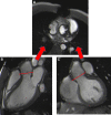
Visualization of the prosthetic orifice of a stentless aortic valve (freestyle) (A) obtained from two orthogonal planes: three-chamber view (B) and coronal LVOT (C). The red line indicates the slice position. Visualization of the leaflets allows easy planimetry.
Figure 4
Figure 4
Visualization of the prosthetic orifice of a surgical stented bioprosthetic aortic valve (saint-jude 25) obtained from a three-chamber view (A). The visualization of leaflets for planimetry is more challenging (B).
Figure 5
Figure 5
Visualization of the orifice of the Edwards-Sapien prosthesis (A) obtained from two orthogonal planes: three-chamber view (B) and coronal LVOT (C). CMR stent-related artefacts prevent accurate aortic valve planimetry.
Figure 6
Figure 6
Example of direct quantification of severe aortic regurgitation for a patient with a degenerated surgical aortic prosthesis (perimount). The perpendicular line (red) above the prosthesis (A) indicates the scanning position for phase-contrast velocity mapping, generating phase (B) and magnitude (C) images and a flow curve image (D). The aortic regurgitant volume is represented by the area under the diastolic flow curve (71 ml); the regurgitant fraction is calculated as the aortic backward volume/aortic forward volume = 71/121.7 = 58.3%.

References

    1. Vahanian A, Beyersdorf F, Praz F, Milojevic M, Baldus S, Bauersachs J, et al. 2021 ESC/EACTS guidelines for the management of valvular heart disease. Eur J Cardiothorac Surg. (2021) 60:727–800. 10.1093/ejcts/ezab389 - DOI - PubMed
    1. Leon MB, Smith CR, Mack MJ, Makkar RR, Svensson LG, Kodali SK, et al. Transcatheter or surgical aortic-valve replacement in intermediate-risk patients. N Engl J Med. (2016) 374:1609–20. 10.1056/NEJMoa1514616 - DOI - PubMed
    1. Reardon MJ, Van Mieghem NM, Popma JJ, Kleiman NS, Søndergaard L, Mumtaz M, et al. Surgical or transcatheter aortic-valve replacement in intermediate-risk patients. N Engl J Med. (2017) 376:1321–31. 10.1056/NEJMoa1700456 - DOI - PubMed
    1. Mack MJ, Leon MB, Thourani VH, Makkar R, Kodali SK, Russo M, et al. Transcatheter aortic-valve replacement with a balloon-expandable valve in low-risk patients. N Engl J Med. (2019) 380:1695–705. 10.1056/NEJMoa1814052 - DOI - PubMed
    1. Popma JJ, Deeb GM, Yakubov SJ, Mumtaz M, Gada H, O’Hair D, et al. Transcatheter aortic-valve replacement with a self-expanding valve in low-risk patients. N Engl J Med. (2019) 380:1706–15. 10.1056/NEJMoa1816885 - DOI - PubMed

LinkOut - more resources