

Primary Cutaneous Adenoid Cystic Carcinoma of the Back: A Case Report
- PMID: 38125236
- PMCID: PMC10731834
- DOI: 10.7759/cureus.49099
Primary Cutaneous Adenoid Cystic Carcinoma of the Back: A Case Report
Abstract
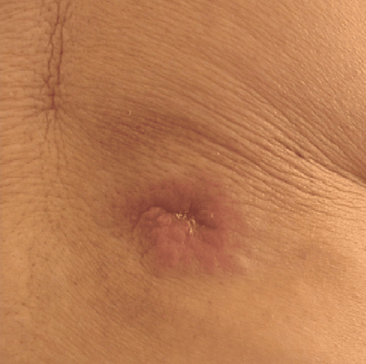
Adenoid cystic carcinoma (ACC) is a rare type of carcinoma that arises from the salivary glands. When ACC is present on the skin with no other primary site of malignancy in the body, it is termed primary cutaneous adenoid cystic carcinoma (PCACC). The only way to differentiate between ACC and other benign cutaneous masses is through the use of histopathology and immunohistochemistry. This case report describes a 67-year-old Asian female with a history of an epidermal inclusion cyst. She was seen in consultation with general surgery for the removal of a mass on her lower back. The initial excision's pathology revealed an ACC with perineural invasion. However, there were positive margins, as the mass was originally thought to be benign. Consequently, she underwent a second procedure for the total excision of the mass, resulting in subsequent negative margins. The patient was referred to radiation oncology; however, she ultimately opted to defer postoperative adjuvant radiation therapy, with the understanding that she would undergo biannual screening examinations.
Keywords: adenoid cystic carcinoma; epidermal inclusion cyst; perineural invasion; radiation therapy; skin cancer.
Copyright © 2023, Dubin et al.
Conflict of interest statement
The authors have declared that no competing interests exist.
Figures

References
-
- Evaluating soft-tissue lumps and bumps. Church DJ, Krumme J, Kotwal S. https://pubmed.ncbi.nlm.nih.gov/30228613/ MO Med. 2017;114:289–294. - PMC - PubMed
-
- Weir CB, St. Hilaire NJ. Treasure Island, FL: StatPearls Publishing; 2023. Epidermal Inclusion Cyst. - PubMed
-
- Zito PM, Scharf R. Treasure Island, FL: StatPearls Publishing; 2023. Epidermoid Cyst. - PubMed
-
- Charifa A, Azmat CE, Badri T. Treasure Island, FL: StatPearls Publishing; 2023. Lipoma Pathology. - PubMed
Publication types
LinkOut - more resources
Full Text Sources