

Paranannizziopsis spp. infections in wild snakes and a qPCR assay for detection of the fungus
- PMID: 38125577
- PMCID: PMC10730940
- DOI: 10.3389/fmicb.2023.1302586
Paranannizziopsis spp. infections in wild snakes and a qPCR assay for detection of the fungus
Abstract
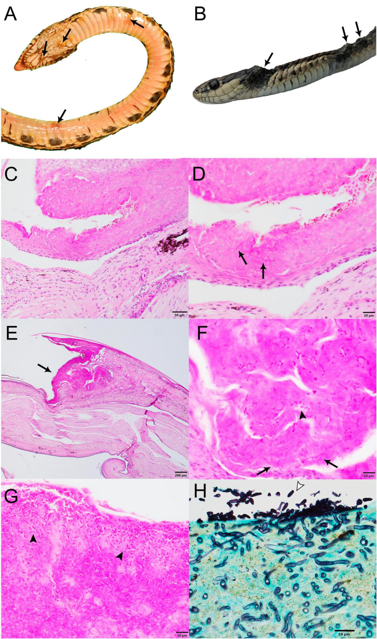
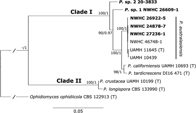
The emergence of ophidiomycosis (or snake fungal disease) in snakes has prompted increased awareness of the potential effects of fungal infections on wild reptile populations. Yet, aside from Ophidiomyces ophidiicola, little is known about other mycoses affecting wild reptiles. The closely related genus Paranannizziopsis has been associated with dermatomycosis in snakes and tuataras in captive collections, and P. australasiensis was recently identified as the cause of skin infections in non-native wild panther chameleons (Furcifer pardalis) in Florida, USA. Here we describe five cases of Paranannizziopsis spp. associated with skin lesions in wild snakes in North America and one additional case from a captive snake from Connecticut, USA. In addition to demonstrating that wild Nearctic snakes can serve as a host for these fungi, we also provide evidence that the genus Paranannizziopsis is widespread in wild snakes, with cases being identified in Louisiana (USA), Minnesota (USA), Virginia (USA), and British Columbia (Canada). Phylogenetic analyses conducted on multiple loci of the fungal strains we isolated identified P. australasiensis in Louisiana and Virginia; the remaining strains from Minnesota and British Columbia did not cluster with any of the described species of Paranannizziopsis, although the strains from British Columbia appear to represent a single lineage. Finally, we designed a pan-Paranannizziopsis real-time PCR assay targeting the internal transcribed spacer region 2. This assay successfully detected DNA of all described species of Paranannizziopsis and the two potentially novel taxa isolated in this study and did not cross-react with closely related fungi or other fungi commonly found on the skin of snakes. The assay was 100% sensitive and specific when screening clinical (skin tissue or skin swab) samples, although full determination of the assay's performance will require additional follow up due to the small number of clinical samples (n = 14 from 11 snakes) available for testing in our study. Nonetheless, the PCR assay can provide an important tool in further investigating the prevalence, distribution, and host range of Paranannizziopsis spp. and facilitate more rapid diagnosis of Paranannizziopsis spp. infections that are otherwise difficult to differentiate from other dermatomycoses.
Keywords: Chrysosporium anamorph of Nannizziopsis vriesii; Onygenales; molecular diagnostics; mycoses; reptile; wildlife disease.
Copyright © 2023 Lorch, Winzeler, Lankton, Raverty, Snyman, Schwantje, Thacker, Knowles, Cai and Grear.
Conflict of interest statement
The authors declare that the research was conducted in the absence of any commercial or financial relationships that could be construed as a potential conflict of interest.
Figures
References
-
- Auliya M. (2003). Hot trade in cool creatures: A review of the live reptile trade in the European Union in the 1990s with a focus on Germany. Brussels: TRAFFIC Europe.
-
- Baker S., Haynes E., Gramhofer M., Stanford K., Bailey S., Christman M., et al. (2019). Case definition and diagnostic testing for snake fungal disease. Herpetol. Rev. 50 279–285.
LinkOut - more resources
Full Text Sources
Miscellaneous