. 2023 Dec 6:14:1287167.
doi: 10.3389/fmicb.2023.1287167. eCollection 2023.

Antiviral functionalization of cellulose using tannic acid and tannin-rich extracts

Affiliations

Antiviral functionalization of cellulose using tannic acid and tannin-rich extracts

Marjo Haapakoski et al. Front Microbiol. .

Abstract

Due to seasonally appearing viruses and several outbreaks and present pandemic, we are surrounded by viruses in our everyday life. In order to reduce viral transmission, functionalized surfaces that inactivate viruses are in large demand. Here the endeavor was to functionalize cellulose-based materials with tannic acid (TA) and tannin-rich extracts by using different binding polymers to prevent viral infectivity of both non-enveloped coxsackievirus B3 (CVB3) and enveloped human coronavirus OC43 (HCoV-OC43). Direct antiviral efficacy of TA and spruce bark extract in solution was measured: EC50 for CVB3 was 0.12 and 8.41 μg/ml and for HCoV-OC43, 78.16 and 95.49 μg/ml, respectively. TA also led to an excellent 5.8- to 7-log reduction of severe acute respiratory syndrome coronavirus 2 (SARS-CoV-2) virus infectivity. TA functionalized materials reduced infectivity already after 5-min treatment at room temperature. All the tested methods to bind TA showed efficacy on paperboard with 0.1 to 1% (w/v) TA concentrations against CVB3 whereas material hydrophobicity decreased activities. Specific signatures for TA and HCoV-OC43 were discovered by Raman spectroscopy and showed clear co-localization on the material. qPCR study suggested efficient binding of CVB3 to the TA functionalized cellulose whereas HCoV-OC43 was flushed out from the surfaces more readily. In conclusion, the produced TA-materials showed efficient and broadly acting antiviral efficacy. Additionally, the co-localization of TA and HCoV-OC43 and strong binding of CVB3 to the functionalized cellulose demonstrates an interaction with the surfaces. The produced antiviral surfaces thus show promise for future use to increase biosafety and biosecurity by reducing pathogen persistence.

Keywords: Raman spectroscopy; antiviral functionalization; bark extract; cellulose; coronaviruses; enteroviruses; tannic acid.


Conflict of interest statement

The authors declare that the research was conducted in the absence of any commercial or financial relationships that could be construed as a potential conflict of interest.

Figures

Figure 1
Figure 1
Dose–response curves for determination of 50% effective concentration (EC50) of tannic acid (TA) and spruce bark extract against viruses and 50% cytotoxic concentration (CC50). Antiviral efficacy of TA (A,E) and spruce bark extract (B,F) was determined against CVB3 (A,B) and HCoV-OC43 (E,F). CVB3 titre in the virus-compound mix was 2 × 107 PFU/ml, while the MOI was 10. HCoV-OC43 titre in the virus-sample mix was 1.6 × 103 PFU/ml and final MOI was 0.008. Toxicity of TA (C,G) and spruce bark extract (D,H) was studied on A549 and MRC-5 cells, respectively. All the experiments were carried out using the CPE inhibition assay. Concentrations of TA and spruce bark extract are represented as Log (10) of μg/ml on the x-axis. The results are expressed as average values ± standard error of the mean (SEM).
Figure 2
Figure 2
Infectivity of HCoV-OC43 and CVB3 on cellulose-based reference materials (A,B) after 5-min incubation at RT and toxicity of materials on cells (C,D). (A) Ten μl of CVB3 (2.0 × 106 PFU/ml) and (B) HCoV-OC43 (9.0 × 106 PFU/ml) were applied on cellulose-based paperboard (P), Sharpcell (SC) and Paptic (P) reference materials (ref) without any additional treatment and CPE assay was exploited to determine the viral infectivity due to treatment. Sample treatments and virus control are normalized against cell control without any infection. Results are presented as average values of 3 biological and 3 technical replicates of each sample ± standard error of the mean (SEM). * p < 0.05, ** p < 0.01, *** p < 0.001 and **** p < 0.0001 versus the virus control (analyzed with one-way ANOVA with Bonferroni test). Toxicity of reference materials was studied on A549 (C) and MRC-5 (D) cells using CPE assay.
Figure 3
Figure 3
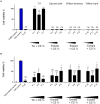
Infectivity of CVB3 and HCoV-OC43 after 5-min incubation on paperboard functionalized with tannic acid (TA) and wood extracts. (A) Ten μl of CVB3 (2.0 × 106 PFU/ml) and (B) HCoV-OC43 (9.0 × 106 PFU/ml) were applied on paperboard materials for 5 min at RT functionalized with TA (P 1–3), spruce bark extract (P 4–6), willow biomass extract (P 7–9) and willow bark extract (P 10–12) in combination with chitosan (CS). Viral infectivity was studied using CPE assay. Increasing amounts of functionalization materials were tested. Detailed chemical composition of each solution used in functionalization is presented in Table 1. Sample treatments and virus control are normalized against cell control without any virus infection. Results are presented as average values of 3 biological and 3 technical replicates of each sample ± standard error of the mean (SEM). * p < 0.05, ** p < 0.01, *** p < 0.001 and **** p < 0.0001 versus the virus control (analyzed with one-way ANOVA with Bonferroni test).
Figure 4
Figure 4
Infectivity of HCoV-OC43 and CVB3 after a 5-min incubation on cellulose-based materials functionalized with tannic acid (TA) and chitosan (CS) or C-PVAm. (A) Ten μl of CVB3 (2.0 × 106 PFU/ml) was incubated for 5 min at RT on reference paperboard (P) and paperboard functionalized with different concentrations of TA and CS (P 13–16) or (B) C-PVAm (P 17–20). (C) Viral infectivity of HCoV-OC43 (9.0 × 105 PFU/ml) was studied on paperboard functionalized with 0.1% TA solution in combination with CS (P 13) and C-PVAm (P 17). (D) Viral infectivity of CVB3 determined after incubation on Paptic (PC) and Sharpcell (SC) materials functionalized with TA and CS. Viral infectivity of (E) CVB3 (2.0 × 106 PFU/ml) and (F) HCoV-OC43 (9.0 × 105 PFU/ml) was determined after 24 h incubation on reference, P13 (0.1% TA) and P15 (1%) paperboard. Efficacy of 5-min treatment was also confirmed at the same time for accurate comparison. Detailed chemical composition of each solution used in functionalization is presented in Table 2. Sample treatments and virus control are normalized against cell control without any virus infection. Results are presented as average values of 3 biological and 3 technical replicates of each sample ± standard error of the mean (SEM). * p < 0.05, ** p < 0.01, *** p < 0.001 and **** p < 0.0001 versus the virus control (analyzed with one-way ANOVA with Bonferroni test).
Figure 5
Figure 5
SMA and C-PAM are tested as binding partners for antiviral functionalization. (A) Ten μl of CVB3 (2.0 × 106 PFU/ml) and (B) HCoV-OC43 (9.0 × 105 PFU/ml) were applied for 5 min at RT on reference paperboard (P) and paperboard functionalized with 1% tannic acid (TA), SMA or C-PAM and with or without chitosan (CS). (C) Viral infectivity of CVB was studied after 5-min incubation on Paptic (PC) material functionalized with 1% TA solution, SMA or C-PAM, and with or without CS. Detailed chemical composition of each solution used in functionalization is presented in Table 3. Sample treatments and virus control are normalized against cell control without any virus infection. Results are presented as average values of 3 biological and 3 technical replicates of each sample ± standard error of the mean (SEM). * p < 0.05, ** p < 0.01, *** p < 0.001 and **** p < 0.0001 versus the virus control (analyzed with one-way ANOVA with Bonferroni test).
Figure 6
Figure 6
Confocal microscopy study of virus treatment on functionalized paperboard for 5 min at RT. After a 5-min treatment of ten μl of CVB3 (8.9 × 108 PFU/ml) and HCoV-OC43 (2.6 × 107 PFU/ml) on three replicates of 1% TA functionalized (P15 in Table 2) and reference paperboard viruses were flushed from the surfaces and applied on cells to determine their infectivity. CVB3 infected A549 cells were labelled for VP1 capsid protein (red) after 5.5 h of infection, while HCoV-OC43 infected MRC-5 cells were labelled for nucleocapsid protein (red) after 15.5 h of infection. DAPI stained cell nuclei are in blue. Scale bars, 30 μm (top) and 50 μm (down). Quantification of the imaging data with CellProfiler is calculated of approximately 650 and 1,276 cells for CVB3 and HCoV-OC43 samples, respectively. * p < 0.05, ** p < 0.01, *** p < 0.001 and **** p < 0.0001 versus the virus control (analyzed with one-way ANOVA with Bonferroni test).
Figure 7
Figure 7
A study on an ability of functionalized material to bind and inactivate viruses. Ten μl of HCoV-OC43 (9.0 × 105 PFU/ml; A,B) and CVB3 (2.0 × 106 PFU/ml; C,D) were applied on reference (Ref) or TA functionalized paperboard (P 15 in Table 2) for 5 min. Viruses were flushed from the surfaces and both the infectivity (A,C) and the amount of viral RNA (B,D) were evaluated of the flushed viruses with CPE and qPCR assays, respectively. qPCR results are presented as a percentage of viral RNA bound to the paperboard materials. Sample treatments and virus control are normalized against cell control without any virus infection. Results are presented as average values of 3 biological and 3 technical replicates of each sample ± standard error of the mean (SEM). * p < 0.05, ** p < 0.01, *** p < 0.001 and **** p < 0.0001 versus the virus control (analyzed with one-way ANOVA with Bonferroni test).
Figure 8
Figure 8
Optical density at 450 nm (OD 405) determined for tannic acid and flushed samples. (A) Different concentrations of TA solution were measured to determine OD 405 that correlates with the amount of TA present. (B) Cellulose materials functionalized with 1% TA were flushed with H2O for 5 min and OD 405 was measured to resolve whether any TA was present in liquid after flushing. Blank value of H2O was subtracted of each OD 405 value measured. Results are presented as average values of 3 replicates of each sample ± standard error of the mean (SEM).
Figure 9
Figure 9
Raman mapping of tannic acid, chitosan and HCoV-OC43 on functionalized material. (A) Raman signature spectra for TA powder and purified HCoV-OC43 on gold surface, as well as for HCoV-OC43 on paperboard with (P 15 in Table 2) and without TA. Three signature peaks for HCoV-OC43 (838, 1,250, and 1,450 cm−1), two signature peaks for TA (1,600 and 1710 cm−1) and one for CS (2,890 cm−1) are indicated in the spectra. (B) Raman mapping of the TA functionalized paperboard with HCoV-OC43 bound for 5 min. An optical image visualizes the area mapped. Mapping of signature peaks of HCoV-OC43 (838 cm−1), TA (1,600 cm−1) and chitosan (2,890 cm−1) from the same area.

References

    1. Abad F. X., Pintó R. M., Bosch A. (1994). Survival of enteric viruses on environmental fomites. Appl. Environ. Microbiol. 60, 3704–3710. doi: 10.1128/aem.60.10.3704-3710.1994, PMID: - DOI - PMC - PubMed
    1. Abdelkhalek A., Qari S. H., Abu-Saied M. A. A.-R., Khalil A. M., Younes H. A., Nehela Y., et al. (2021). Chitosan nanoparticles inactivate alfalfa mosaic virus replication and boost innate immunity in Nicotiana glutinosa plants. Plants 10:2701. doi: 10.3390/plants10122701, PMID: - DOI - PMC - PubMed
    1. Alnuqaydan A. M., Almutary A. G., Sukamaran A., Yang B. T. W., Lee X. T., Lim W. X., et al. (2021). Middle East respiratory syndrome (MERS) virus—pathophysiological Axis and the current treatment strategies. AAPS PharmSciTech 22:173. doi: 10.1208/s12249-021-02062-2, PMID: - DOI - PMC - PubMed
    1. Amankwaah C., Li J., Lee J., Pascall M. A. (2020). Development of antiviral and bacteriostatic chitosan-based food packaging material with grape seed extract for murine norovirus, Escherichia coli and Listeria innocua control. Food Sci. Nutr. 8, 6174–6181. doi: 10.1002/fsn3.1910 - DOI - PMC - PubMed
    1. Anany H., Chen W., Pelton R., Griffiths M. W. (2011). Biocontrol of listeria monocytogenes and Escherichia coli O157:H7 in meat by using phages immobilized on modified cellulose membranes. Appl. Environ. Microbiol. 77, 6379–6387. doi: 10.1128/AEM.05493-11, PMID: - DOI - PMC - PubMed