Review
. 2023 Dec;19(4):230140.
doi: 10.1183/20734735.0140-2023. Epub 2023 Dec 19.

A practical approach to the diagnosis and management of malignant pleural effusions in resource-constrained settings

Affiliations
Review

A practical approach to the diagnosis and management of malignant pleural effusions in resource-constrained settings

Jane A Shaw et al. Breathe (Sheff). 2023 Dec.

Abstract

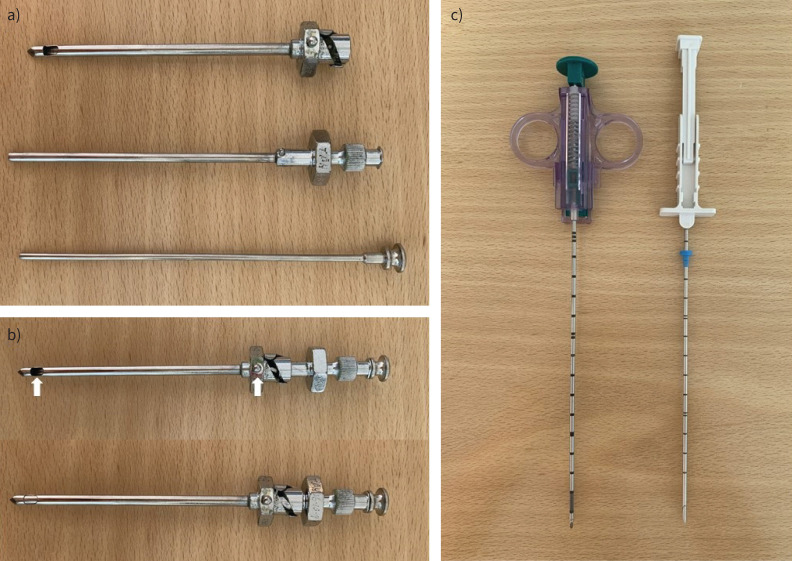
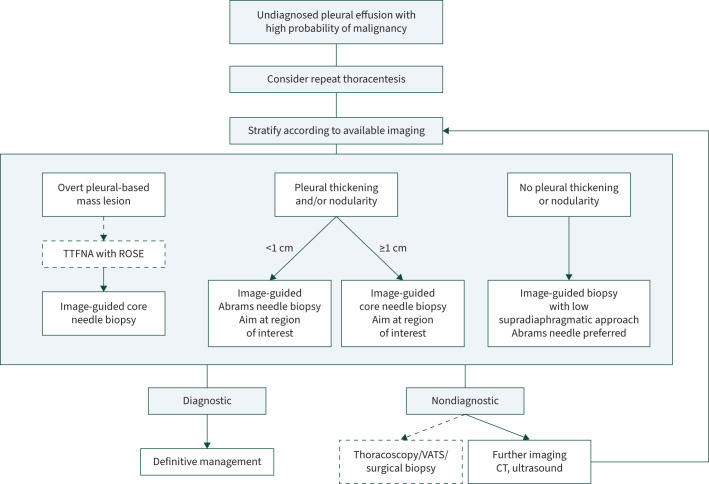
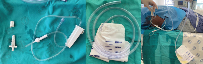
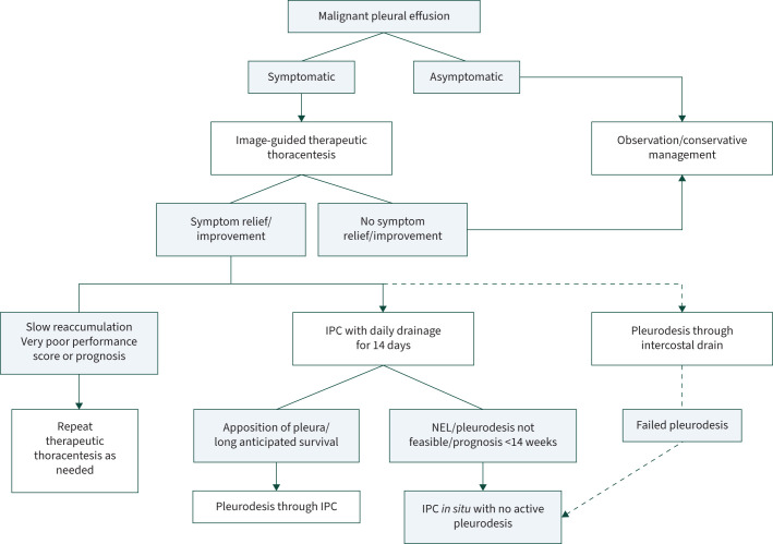
No pleural intervention in a patient with confirmed malignant pleural effusion (MPE) prolongs life, but even the recommended interventions for diagnosis and palliation can be costly and therefore unavailable in large parts of the world. However, there is good evidence to guide clinicians working in low- and middle-income countries on the most cost-effective and clinically effective strategies for the diagnosis and management of MPE. Transthoracic ultrasound-guided closed pleural biopsy is a safe method of pleural biopsy with a diagnostic yield approaching that of thoracoscopy. With the use of pleural fluid cytology and ultrasound-guided biopsy, ≥90% of cases can be diagnosed. Cases with an associated mass lesion are best suited to an ultrasound-guided fine needle aspiration with/without core needle biopsy. Those with diffuse pleural thickening and/or nodularity should have an Abrams needle (<1 cm thickening) or core needle (≥1 cm thickening) biopsy of the area of interest. Those with insignificant pleural thickening should have an ultrasound-guided Abrams needle biopsy close to the diaphragm. The goals of management are to alleviate dyspnoea, prevent re-accumulation of the pleural effusion and minimise re-admissions to hospital. As the most cost-effective strategy, we suggest early use of indwelling pleural catheters with daily drainage for 14 days, followed by talc pleurodesis if the lung expands. The insertion of an intercostal drain with talc slurry is an alternative strategy which is noninferior to thoracoscopy with talc poudrage.

Educational aims: To provide clinicians practising in resource-constrained settings with a practical evidence-based approach to the diagnosis and management of malignant pleural effusions.To explain how to perform an ultrasound-guided closed pleural biopsy.To explain the cost-effective use of indwelling pleural catheters.


Conflict of interest statement

Conflict of interest: J.A. Shaw reports receiving an honorarium from AstraZeneca, outside the submitted work. E.H. Louw has nothing to disclose. C.F.N. Koegelenberg reports receiving an honorarium from GSK and AstraZeneca, outside the submitted work.

Figures

FIGURE 1
FIGURE 1
Typical imaging findings in a malignant pleural effusion. a) A chest radiograph of a large right-sided malignant pleural effusion with an associated mass in the right upper zone; b) an axial slice from thoracic computed tomography (CT) in a patient with a large right-sided malignant pleural effusion, showing a thickened abnormal pleural with a distinct pleural-based mass lesion amenable to biopsy (arrow); c) a thoracic ultrasound image of large malignant effusion (E) with a markedly thickened parietal pleura (pleural thickening (PT)). Atelectatic lung (L) is seen in the effusion, and the highly echogenic diaphragmatic pleura (D) on the right.
FIGURE 2
FIGURE 2
Needles for image-guided closed pleural biopsy. a) The traditional reusable Abrams pleural biopsy needle; the three parts of the needle are shown (from top to bottom): the outer cylinder, the inner cylinder with cutting edge, and the stylet. b) The assembled Abrams needle in the open position with the notch indicator on the grip in line with the specimen open notch (arrows). Below this is the needle in the closed position, as the inner cylinder has been rotated down into the outer cylinder. c) Typical core-cutting needles: on the left, an automated needle which will trigger a closing action after being primed; on the right, a manual needle.
FIGURE 3
FIGURE 3
A practical approach to the diagnosis of a malignant pleural effusion in a severely resource-constrained setting. Transthoracic fine-needle aspiration (TTFNA) with rapid on-site evaluation (ROSE) by a pathologist may not be available, in which case an image-guided biopsy would the first investigation after thoracentesis in an effusion with an overt pleural-based mass lesion. Where thoracoscopy, video-assisted thoracoscopic surgery (VATS) and surgical biopsy are unavailable, then we suggest obtaining either computed tomography (CT) or ultrasound, depending on what has already been done. Then stratify according to this imaging and repeat the biopsy with a new target. Figure created using BioRender.com.
FIGURE 4
FIGURE 4
An indwelling pleural catheter (IPC) with alternative drainage option. Shown is the equipment supplied in the typical IPC pack for attachment to the single-use drainage bottles, and how these can be attached to a conventional urine catheter bag instead.
FIGURE 5
FIGURE 5
A practical approach to the management of a malignant pleural effusion in a severely resource constrained setting. Where the patient experiences and improvement in symptoms after thoracentesis and has a reasonable prognosis, an indwelling pleural catheter (IPC) is the preferred intervention. Talc pleurodesis may be done through the IPC, unless there is a nonexpandable lung (NEL), or the life expectancy is <14 weeks. If no IPC is available, an intercostal drain with talc pleurodesis is an acceptable alternative. Figure created using BioRender.com.

Comment in

  • doi: 10.1183/20734735.0230-2023

References

    1. Rodrîguez-Panadero F, Borderas Naranjo F, López Mejîas J. Pleural metastatic tumours and effusions. Frequency and pathogenic mechanisms in a post-mortem series. Eur Respir J 1989; 2: 366–369. doi:10.1183/09031936.93.02040366 - DOI - PubMed
    1. Adeoye PO, Johnson WR, Desalu OO, et al. . Etiology, clinical characteristics, and management of pleural effusion in Ilorin, Nigeria. Niger Med J 2017; 58: 76–80. doi:10.4103/0300-1652.219349 - DOI - PMC - PubMed
    1. Ebrahimi H, Aryan Z, Saeedi Moghaddam S, et al. . Global, regional, and national burden of respiratory tract cancers and associated risk factors from 1990 to 2019: a systematic analysis for the Global Burden of Disease Study 2019. Lancet Respir Med 2021; 9: 1030–1049. doi:10.1016/S2213-2600(21)00164-8 - DOI - PMC - PubMed
    1. Koegelenberg CFN, Bennji SM, Boer E, et al. . The current aetiology of malignant pleural effusion in the Western Cape Province, South Africa. S Afr Med J 2018; 108: 275–277. doi:10.7196/SAMJ.2018.v108i4.12936 - DOI - PubMed
    1. Roberts ME, Neville E, Berrisford RG, et al. . Management of a malignant pleural effusion: British Thoracic Society pleural disease guideline 2010. Thorax 2010; 65: Suppl. 2, ii32–ii40. doi: 10.1136/thx.2010.136994 - DOI - PubMed

LinkOut - more resources