

doi: 10.1183/20734735.0153-2023.
Epub 2023 Dec 19.
Thoracic ultrasound for pneumothorax and infectious effusion: from equine beginnings to clinical cornerstone
Affiliations
- PMID: 38125807
- PMCID: PMC10729822
- DOI: 10.1183/20734735.0153-2023
Item in Clipboard
Thoracic ultrasound for pneumothorax and infectious effusion: from equine beginnings to clinical cornerstone
Breathe (Sheff).
2023 Dec.
Abstract
Routine clinical application of thoracic ultrasound has greatly enhanced the process of diagnosing and treating patients with pneumothorax and infectious effusion by minimising radiation exposure and facilitating prompt diagnosis https://bit.ly/3FO6jBg.
Copyright ©ERS 2023.
Conflict of interest statement
Conflicts of interest: C. Falster reports personal fees from Bristol-Myers Squibb, outside the submitted work. T. Gille reports personal fees from Roche S.A.S., and other support from Oxyvie (oxygen provider), Vivisol France (oxygen provider) and Menarini France, outside the submitted work; T. Gille is a current member of the Breathe editorial board. The remaining authors have nothing to disclose.
Figures

a) A horse is subjected to a computed tomography scan. Evidently, this is a resource- and time-consuming procedure requiring a broad array of veterinary competencies and staff. Image from IMV Imaging; reproduced with permission. b) A horse is examined using ultrasound. Note the calm body language of the horse, reflecting the modest discomfort experienced during the procedure. Image from Marion duPont Scott Equine Medical Center; reproduced with permission.
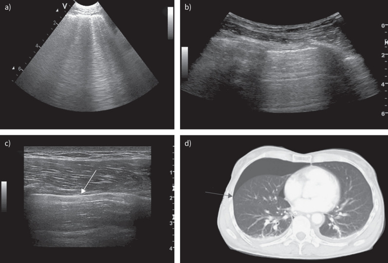
a) Multiple B-lines, ruling out the presence of pneumothorax in the scanned zone. The B-lines are hyperechoic, go all the way to the bottom of the screen, erase horizontal A-lines and move with lung sliding. b) A normal standard view with lung sliding (unsurprisingly, this finding is not visible on a still image). c) A lung-point. Lung sliding is present at the left side of the image, whereas the pleural line is completely static on the right side. The white arrow indicates the lung point, which represents the junction where the lung ends and the pneumothorax starts. d) A computed tomography scan from the same patient as in c); the arrow corresponds to the lung point.

a) A simple pleural effusion. The fluid is homogeneous, hypoechoic and devoid of septation. To the right of the image, the diaphragm is seen; beneath it, the liver. b) A simple parapneumonic effusion. The pneumonic tissue is hypoechoic compared to adjacent structures and contains air bronchograms, small hyperechoic areas, representing air in the bronchi. c) A complicated parapneumonic effusion (Eff) with septation spanning from the diaphragm above the liver (Lvr) to the pneumonic lung (Pnm). d) Pleural effusion characterised by comprehensive fibrinous material, highly suggestive of empyema.
Comment in
-
Diagnosing and managing pleural disease.Breathe (Sheff). 2023 Dec;19(4):230230. doi: 10.1183/20734735.0230-2023. Epub 2023 Dec 19. Breathe (Sheff). 2023. PMID: 38127543 Free PMC article.
References
-
- Lichtenstein D. Lung ultrasound in the critically ill. In: Vincent JL, ed. Yearbook of Intensive Care and Emergency Medicine. Heidelberg, Springer, 2004; pp. 625–644.
LinkOut - more resources
Full Text Sources