

Added value of 3D THRIVE (T1-weighted high-resolution isotropic volume examination) MRI pulse sequence in the detection of bony erosions of sacroiliac joints in patients of spondyloarthritis
- PMID: 38125811
- PMCID: PMC10731439
- DOI: 10.5114/pjr.2023.132877
Added value of 3D THRIVE (T1-weighted high-resolution isotropic volume examination) MRI pulse sequence in the detection of bony erosions of sacroiliac joints in patients of spondyloarthritis
Abstract
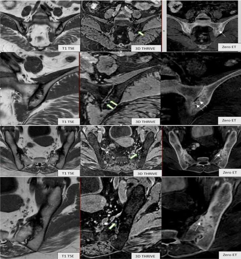
Purpose: Early depiction of bony erosions in sacroiliac (SI) joints increases the diagnostic accuracy of spondyloarthritis. The new 3D magnetic resonance imaging (MRI) sequence THRIVE (T1-weighted high-resolution isotropic volume examination) can depict cartilage erosions in sacroiliac joints. The aim of the study was to compare the diagnostic capacity of the new MRI sequence 3D THRIVE (T1-weighted high-resolution isotropic volume examination) with the routinely used T1 TSE pulse sequence in the depiction of structural erosions in sacroiliac joints by using MRI sequence zero echo time (zero ET) as a reference standard.
Material and methods: Seventy five adult patients were included in this study. They underwent MRI sacroiliac joints examination using routine T1 TSE and STIR pulse sequences with the addition of the new 3D THRIVE and zero echo time (zero ET) sequences. Images of T1 TSE, 3D THRIVE, and zero ET sequences were evaluated by 2 radiolo-gists separately for the detection of sacroiliac joints erosions, then a comparison between T1 TSE and 3D THRIVE sequences was done using a CT-like image MRI sequence zero ET as a reference standard. Sensitivity, specificity, and accuracy for each sequence were calculated by the 2 readers.
Results: Sensitivity, specificity, and accuracy of 3D THRIVE were higher than those of T1 TSE for reader 1 (sensitivity: 94.5% vs. 86.2%; specificity: 93.4% vs. 85.1%; and accuracy 95.2% vs. 88.5%) and for reader 2 (sensitivity: 93.3% vs. 79.9%; specificity: 94.7% vs. 86.2%; and accuracy 95.8% vs. 82.1%).
Conclusions: Using CT-like image MRI sequence zero ET as the reference standard, 3D THRIVE pulse sequencing of the sacroiliac joints has much better diagnostic value in the depiction of bony erosions in patients suspected having spondyloarthritis as compared to the routinely used T1 TSE sequence.
Keywords: T1 turbo spin echo; T1-weighted high-resolution isotropic volume examination; magnetic resonance imaging; sacroiliac joints.
© Pol J Radiol 2023.
Conflict of interest statement
The authors report no conflict of interest.
Figures


References
-
- Sieper J, Rudwaleit M, Baraliakos X, et al. . The Assessment of Spondyloarthritis International Society (ASAS) handbook: a guide to assess spondyloarthritis. Ann Rheum Dis 2009; 68 Suppl 2: ii1-ii44. - PubMed
-
- Rudwaleit M, Jurik AG, Hermann KG, et al. . Defining active sacroiliitis on magnetic resonance imaging (MRI) for classification of axial spondyloarthritis: a consensual approach by the ASAS/OMERACT MRI group. Ann Rheum Dis 2009; 68: 1520-1527. - PubMed
-
- Mandl P, Navarro-Compan V, Terslev L, et al. . EULAR recommendations for the use of imaging in the diagnosis and management of spondyloarthritis in clinical practice. Ann Rheum Dis 2015; 74: 1327-1339. - PubMed
-
- van den Berg R, de Hooge M, Rudwaleit M, et al. . ASAS modification of the Berlin algorithm for diagnosing axial spondyloarthritis: results from the Spondyloarthritis Caught Early (SPACE)-cohort and from the Assessment of Spondyloarthritis International Society (ASAS)-cohort. Ann Rheum Dis 2013; 72: 1646-1653. - PubMed
-
- Weber U, Ostegaard M, Lambert RGW, et al. . Candidate lesion-based criteria for defining a positive sacroiliac joint MRI in two cohorts of patients with axial spondyloarthritis. Ann Rheum Dis 2015; 74: 1976-1982. - PubMed
LinkOut - more resources
Full Text Sources