

Understanding the diagnosis of catheter-related bloodstream infection: real-time monitoring of biofilm growth dynamics using time-lapse optical microscopy
- PMID: 38125909
- PMCID: PMC10731284
- DOI: 10.3389/fcimb.2023.1286527
Understanding the diagnosis of catheter-related bloodstream infection: real-time monitoring of biofilm growth dynamics using time-lapse optical microscopy
Abstract
Background: The differential time to positivity (DTTP) technique is recommended for the conservative diagnosis of catheter-related bloodstream infection (C-RBSI). The technique is based on a 120-minute difference between microbial growth in blood drawn through the catheter and blood drawn through a peripheral vein. However, this cut-off has failed to confirm C-RBSI caused by Candida spp. and Staphylococcus aureus.
Objective: We hypothesized that the biofilm of both microorganisms disperses faster than that of other microorganisms and that microbial load is rapidly equalized between catheter and peripheral blood. Therefore, our aim was to compare the biofilm dynamics of various microorganisms.
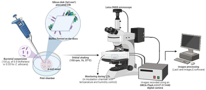
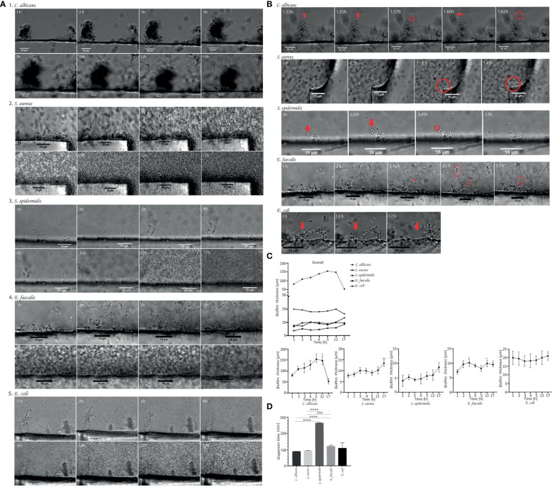
Methods: Biofilm of ATCC strains of methicillin-resistant Staphylococcus epidermidis, methicillin-susceptible S. aureus, Enterococcus faecalis, Escherichia coli and Candida albicans was grown on silicon disks and analyzed using time-lapse optical microscopy. The time-lapse images of biofilms were processed using ImageJ2 software. Cell dispersal time and biofilm thickness were calculated.
Results: The mean (standard deviation) dispersal time in C. albicans and S. aureus biofilms was at least nearly 3 hours lower than in biofilm of S. epidermidis, and at least 15 minutes than in E. faecalis and E. coli biofilms.
Conclusion: Our findings could explain why early dissemination of cells in C. albicans and S. aureus prevents us from confirming or ruling out the catheter as the source of the bloodstream infection using the cut-off of 120 minutes in the DTTP technique. In addition, DTTP may not be sufficiently reliable for E. coli since their dispersion time is less than the cut-off of 120 minutes.
Keywords: biofilm; catheter-related bloodstream infections; differential time to positivity; growth; time-lapse optical microscopy.
Copyright © 2023 Díaz-Navarro, Samaniego, Piqueras, Díez, Hafian, Manzano, Muñoz and Guembe.
Conflict of interest statement
The authors declare that the research was conducted in the absence of any commercial or financial relationships that could be construed as a potential conflict of interest.
Figures
Similar articles
-
Evaluation of Antimicrobial Durability and Anti-Biofilm Effects in Urinary Catheters Against Enterococcus faecalis Clinical Isolates and Reference Strains.Balkan Med J. 2017 Dec 1;34(6):546-552. doi: 10.4274/balkanmedj.2016.1853. Balkan Med J. 2017. PMID: 29215338 Free PMC article.
-
Comparison of linezolid and vancomycin lock solutions with and without heparin against biofilm-producing bacteria.Am J Health Syst Pharm. 2017 May 1;74(9):e193-e201. doi: 10.2146/ajhp150804. Am J Health Syst Pharm. 2017. PMID: 28438824
-
Caspofungin Inhibits Mixed Biofilms of Candida albicans and Methicillin-Resistant Staphylococcus aureus and Displays Effectiveness in Coinfected Galleria mellonella Larvae.Microbiol Spectr. 2021 Oct 31;9(2):e0074421. doi: 10.1128/Spectrum.00744-21. Epub 2021 Oct 13. Microbiol Spectr. 2021. PMID: 34643410 Free PMC article.
-
Tetrasodium EDTA as a novel central venous catheter lock solution against biofilm.Infect Control Hosp Epidemiol. 2005 Jun;26(6):515-9. doi: 10.1086/502577. Infect Control Hosp Epidemiol. 2005. PMID: 16018425
-
High success rate in salvage of catheter-related bloodstream infections due to Staphylococcus aureus, on behalf of project group of Italian society of nephrology.J Vasc Access. 2020 May;21(3):336-341. doi: 10.1177/1129729819875323. Epub 2019 Sep 12. J Vasc Access. 2020. PMID: 31512986 Review.
Cited by
-
Vascular catheter-related infections: an endemic disease in healthcare institutions. An opinion paper of the Spanish Society of Cardiovascular Infections (SEICAV).Rev Esp Quimioter. 2024 Oct;37(5):387-400. doi: 10.37201/req/051.2024. Epub 2024 Jun 26. Rev Esp Quimioter. 2024. PMID: 38916720 Free PMC article. Review.
-
Reliability of differential time to positivity technique for diagnosing catheter-related bloodstream infections: a retrospective analysis.Microbiol Spectr. 2025 Jun 3;13(6):e0267824. doi: 10.1128/spectrum.02678-24. Epub 2025 Apr 8. Microbiol Spectr. 2025. PMID: 40197990 Free PMC article.
-
The influence of marine fungal meroterpenoid meroantarctine A toward HaCaT keratinocytes infected with Staphylococcus aureus.J Antibiot (Tokyo). 2024 Dec;77(12):812-822. doi: 10.1038/s41429-024-00771-x. Epub 2024 Sep 10. J Antibiot (Tokyo). 2024. PMID: 39256545
References
-
- Bouza E., Alcalá L., Muñoz P., Martín-Rabadán P., Guembe M., Rodríguez-Créixems M. (2013). Can microbiologists help to assess catheter involvement in candidaemic patients before removal? Clin. Microbiol. infection Off. Publ. Eur. Soc. Clin. Microbiol. Infect. Dis. 19 (2), E129–E135. doi: 10.1111/1469-0691.12096 - DOI - PubMed
-
- Bouzidi H., Emirian A., Marty A., Chachaty E., Laplanche A., Gachot B., et al. . (2018). Differential time to positivity of central and peripheral blood cultures is inaccurate for the diagnosis of Staphylococcus aureus long-term catheter-related sepsis. J. Hosp. infection 99 (2), 192–199. doi: 10.1016/j.jhin.2018.01.010 - DOI - PubMed
Publication types
MeSH terms
LinkOut - more resources
Full Text Sources
Medical
Miscellaneous